product description
Propidium iodide (PI) is a commonly used nuclear fluorescent dye, which is an analog of ethidium bromide (EB). It can be embedded between DNA base pairs and bind without significant sequence preference, binding to approximately one dye molecule every 4-5 base pairs. PI can also bind to RNA, and to distinguish DNA from RNA staining, nucleases are usually added for treatment. The maximum excitation/emission wavelength of PI aqueous solution is 493/636 nm, but after binding with nucleic acid, the fluorescence signal is enhanced by 20-30 times, and the maximum excitation/emission wavelength becomes 535/617 nm. PI is suitable for fluorescence microscopy, confocal microscopy, flow cytometry, and fluorescence analysis.
PI cannot penetrate the cell membrane and is excluded from living cells, but can penetrate damaged cell membranes and stain the nucleus. By utilizing this characteristic, it is usually used in conjunction with live cell fluorescent probes such as Calcein AM, Hoechst 33258 (item number: AC12L012), or Hoechst 33342 (item number: AC12L022), for staining and identification of both live and dead cells, for research related to cell apoptosis. Meanwhile, PI is used as a counterstain for multiplex fluorescent staining, compatible with various cell labeling techniques, including direct or indirect fluorescent antibody detection, mRNA in situ hybridization, cell structure specific fluorescent probe detection, and tissue staining. PI staining is also applicable for cell cycle analysis.
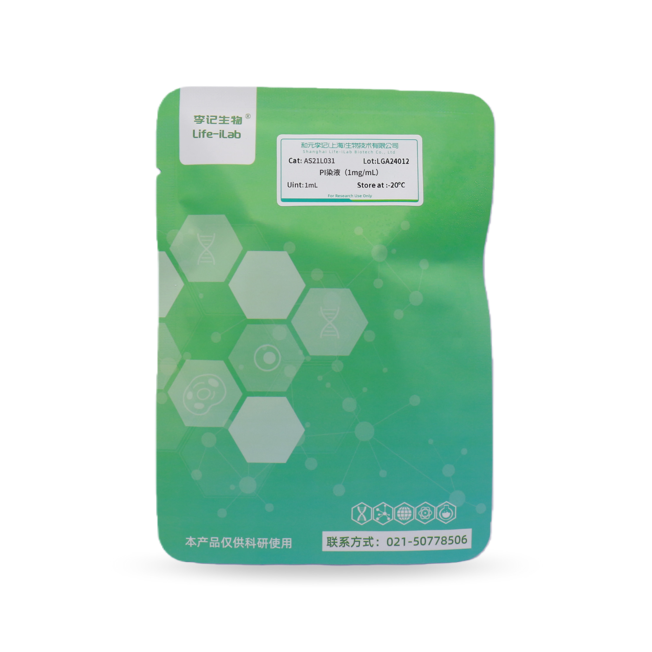
This product is a PI storage solution in the form of an aqueous solution, with a concentration of 1 mg/ml. It can be diluted to the appropriate working concentration before use.
ordering information
product name | Item number | specifications |
PI staining solution (1mg/mL) | AS21L031 | 1mL |
Transportation and storage
Blue ice transportation- Store at 20 ℃ away from light, with a shelf life of 6 months.
Usage
1. Staining steps for adherent cells (fluorescence microscopy detection)
1.1 Sample preparation: Choose appropriate steps to fix cells based on your own sample. PI staining is usually performed after other staining is completed. PI counterstaining requires cells to undergo permeabilization treatment.
1.2 RNase enzyme treatment: If the sample is fixed with polyformaldehyde, formaldehyde, or glutaraldehyde, RNase treatment is required. If the sample is fixed with methanol/acetic acid or acetone, this step is usually not required.
1.2.1 Equilibrate the sample in a solution of 2 × SSC (0.3 M NaCl, 0.03 M sodium citrate, pH 7.0);
1.2.2 Incubate this product in a 2 × SSC solution containing 100 µ g/mL DNase free RNase at 37 ° C for 20 minutes;
1.2.3 Clean the sample 3 times with 2 × SSC solution, each time for 1 minute.
1.3 Re dyeing
1.3.1 2 × SSC (0.3 M NaCl, 0.03 M sodium citrate, pH 7.0) solution equilibrium sample;
1.3.2 Dilute 1mg/ml (1.5 mM) PI storage solution 1:3000 directly with 2 × SSC to obtain 500 nM PI working solution. Usually, adding 300 µ l of staining solution is sufficient for one cover glass cell slide, staining for 1-5 minutes.
1.3.3 Clean 2 × SSC several times, drain excess buffer solution, and add anti quenching agent to seal the sheet.
1.3.4 Select appropriate filters for fluorescence microscopy for observation.
2. Suspension cell counterstaining steps (flow cytometry detection)
2.1 Sample preparation: Choose appropriate steps to fix cells based on your own sample. Alternatively, use the following steps:
Collect a certain amount of cells with a density of approximately 2 × 10⁵~1 × 10⁶. Collect cells by centrifugation, aspirate the supernatant, and gently tap the tube wall with your hand to resuspend the remaining liquid. Then add 1ml of PBS stored at room temperature; Transfer all resuspended cells to 4ml of anhydrous ethanol pre cooled at -20 º C, and slowly add the cell suspension to the ethanol while high-speed vortexing and mixing. Fix in ethanol at -20 º C for 5-15 minutes. Collect cells by centrifugation and remove ethanol. Gently tap the tube wall with your hand to loosen the cells, then add 5ml of room temperature PBS. Allow cells to hydrate for 15 minutes;
2.2 Re dyeing
Using staining solution (100mM Tris, Dilute 1mg/ml (1.5 mM) PI storage solution 1:500 with pH 7.4, 150mM NaCl, 1mM CaCl2, 0.5mM MgCl2, 0.1% NP-40 to obtain a 3 µ M PI working solution. 1ml of PI staining solution is sufficient for the detection of each cell sample.
Note: The concentration of the working solution can be adjusted according to the experimental system, or the PI storage solution can be directly diluted to the desired concentration using buffer solutions such as PBS and HBSS. After the final step of sample preparation, collect the cells by centrifugation, remove the supernatant, gently tap the tube wall with your hand to loosen the cells, and add 1ml of PI staining working solution. After incubating at room temperature for 15 minutes, flow cytometry was used for cell analysis. If observed under a flow microscope, the sample needs to be centrifuged, the supernatant removed, and the cells resuspended in fresh buffer. Take 1 drop of suspension onto a glass slide, cover it with a cover slip, and observe.
3. Chromatin FISH counterstaining steps
3.1 Sample Preparation: Prepare samples according to standard procedures. The final step before counterstaining is to wash the sample with deionized water to remove any residual buffer salts on the slide. Dry at room temperature. This step helps reduce non-specific background staining.
3.2 Re dyeing
Preparation of working solution: Dilute 1mg/ml (1.5mM) of PI storage solution 1:1000 directly with PBS buffer to obtain a 1.5 µ M PI staining working solution. Add 300 µ L of working fluid directly to the sample. If necessary, add freshly prepared RNase A (final concentration: 10 mg/mL) to the working solution. Plastic cover glass can be used to evenly distribute the dye solution on the glass slide. Incubate the sample at room temperature in the dark for 30 minutes; If RNase is added, incubate at 37 º C. Remove the cover glass and wash it with PBS or deionized water to remove unbound dyes; Use absorbent paper towel to absorb residual liquid around the sample, cover the glass cover slide and seal the edge of the cover slide with paraffin or nail polish. Anti fluorescence quenching agents can also be used for film sealing. Select appropriate filters for fluorescence microscopy for observation.
matters needing attention
1. This product is only for scientific experimental research and should not be used in clinical diagnosis, treatment, or other fields.
2. For your safety and health, please wear lab clothes and disposable gloves when operating.
3. Propidium iodide (PI) is a known mutagen, so the PI solution needs to be treated with activated carbon before disposal.
4. It is recommended to take 1-2 samples for preliminary experiments when using this kit for the first time.
Related product recommendations
Universal tissue cell fixative (4% paraformaldehyde) (item number: AC28L112)
Anti fluorescence quencher (item number: AC28L512)
Hoechst 33258 (item number: AC12L012)
Hoechst 33342 (item number: AC12L022)
1X PBS (sterile) (item number: AC08L011)
This product is only for scientific experimental research and should not be used in clinical diagnosis, treatment, or other fields.


 Back
Back
 Back
Back


