product description
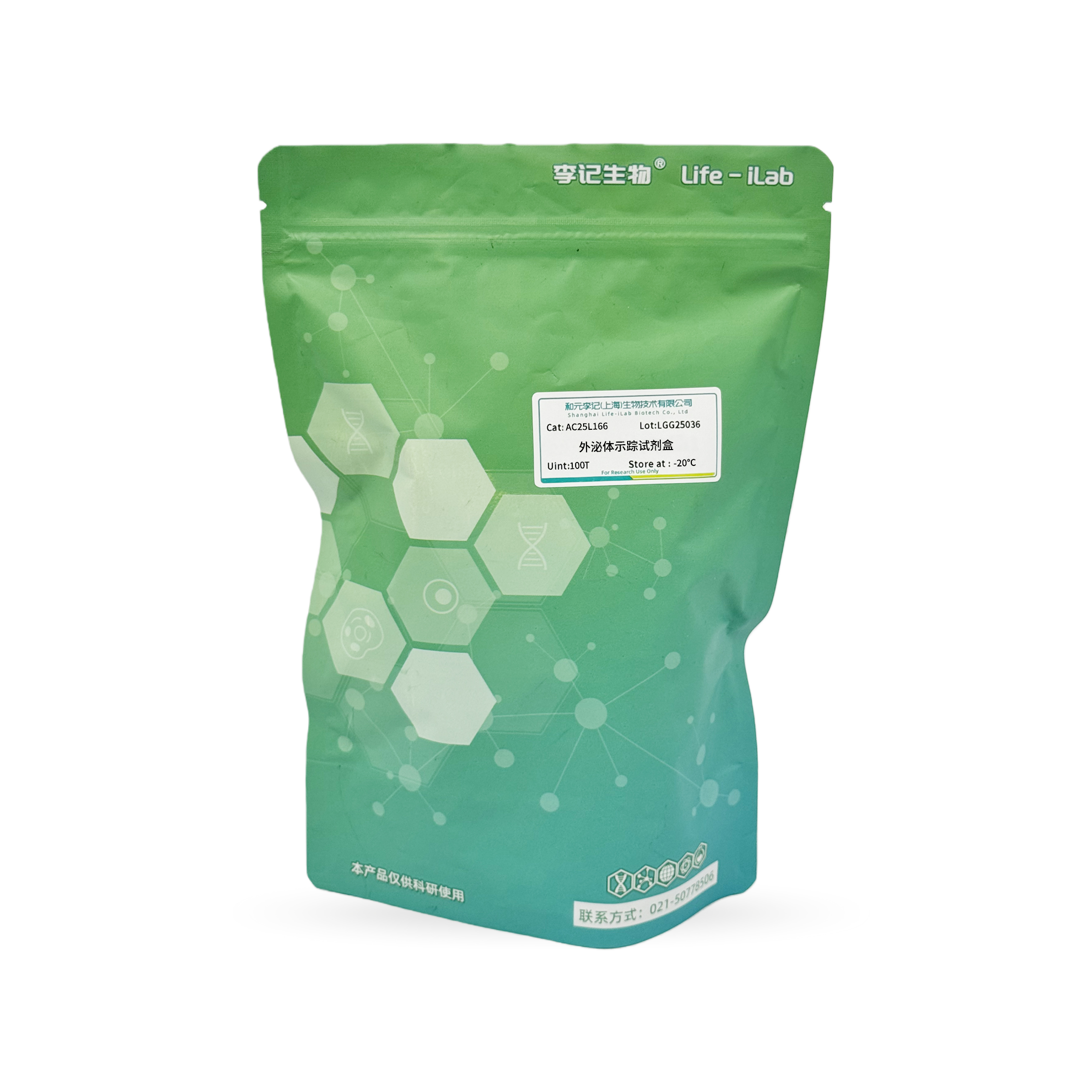
This kit contains an innovative Exosome Tracker, a tracer dye probe specifically designed for fluorescent labeling of extracellular vesicles. The labeling efficiency of Exosome Tracker is higher than other probes, and it adopts a double-layer membrane structure embedded covalently in extracellular vesicles, which is superior to lipophilic or hydrophobic dyes, avoiding signal interference caused by self-assembled nanoparticles and thus improving labeling efficiency. Exosome Tracker can easily and effectively track and monitor extracellular vesicles in vitro, as it has a long half-life in vivo. The labeled extracellular vesicles can be used for animal in vivo tracking experiments. The excitation wavelength is 580 nm and the emission wavelength is 664 nm.
ordering information
product name | Item number | specifications |
Extracellular vesicle tracer kit | AC25L166 | 100T |
Product components
components | specifications |
A. Exosome Tracker | 100μL |
B. Exosome Tracker Remover | 1mL |
Transportation and storage
Blue ice transportation- Store at 20 ℃ away from light, with a shelf life of 12 months.
Usage
Self provided reagents and consumables: DAPI, PBS (freshly prepared, sterile), confocal petri dishes, buffer replacement kits, or other buffer replacement products.
1. Preparation of reagents
Exosome Tracker needs to be removed from -20 ℃ and thawed in the dark before use. Note: Exosome Tracker has a concentration of 200 x, it is recommended to pack and store in the dark at -20 ℃ upon receipt.
2. Marking
(1) Labeling extracellular vesicles: Add Exosome Tracker to the extracellular vesicle solution at a ratio of 1:200 (v/v), incubate at 37 ℃ in the dark for 1 hour, gently invert up and down to ensure uniform staining.
(2) Remove free Exosome Tracker: Add 0.1 times the volume of Exosome Tracker Remover, incubate at room temperature in the dark for 10 minutes to neutralize excess probes, and then replace the solution with PBS using a buffer replacement kit to remove free Exosome Tracker and obtain pure fluorescently labeled exosomes. Note: Choose the appropriate buffer replacement product based on the sample volume: S-buffer replacement kit (item number: AC25L491) suitable for 100-180 μ L; M-buffer replacement kit (item number: AC25L492) is suitable for 0.2-0.5 mL; The L-buffer replacement kit (item number: AC25L493) is suitable for 1-2.5 mL.
3. Tracer
For cell experiments
(1) Inoculate cells: Inoculate cells into confocal specialized petri dishes at an appropriate density for cultivation.
(2) Incubate extracellular vesicles: When the cell grows to 70%, add fluorescently labeled extracellular vesicles and incubate in the dark under cell culture conditions. Note: The specific incubation time is determined based on the cell type and extracellular vesicle source, usually 2 hours, with an adjustable range of 2-4 hours.
(3) Cleaning and staining: Remove the petri dish, wash it three times with PBS, add DAPI solution, incubate at room temperature in the dark for 10 minutes, and then wash it three times with PBS. If detected directly, the fixed step can be skipped, otherwise fix the cells with 4% paraformaldehyde.
(4) Fluorescence detection: Observe cells and extracellular vesicles under a laser confocal microscope, and take photos for recording.
For animal experiments
(1) Preparation of experimental animals: For example, select 6-8 week old mice or other appropriate animal models and administer a certain dose of extracellular vesicles twice a week.
(2) Extracellular vesicle administration: Inject exosomes labeled with Exosome Tracker into animals according to the predetermined dosage, administration method, and frequency of exosomes.
(3) Detection: After administration of extracellular vesicles, in vivo luminescence imaging or other fluorescence detection is usually performed 6-8 weeks later.
precautions
1. This product is only for scientific experimental research and should not be used in clinical diagnosis, treatment, or other fields.
2. For your safety and health, please wear lab clothes and disposable gloves when operating.
3. Fluorescent dyes all have quenching problems. Please avoid light during operation to slow down fluorescence quenching
4. This product needs to explore the optimal working concentration and staining time based on cell type, culture conditions, and application direction.
Related product recommendations
Extracellular vesicle isolation kit (SEC method) (item number: AC25L434)
Extracellular vesicle isolation kit (magnetic bead method) (item number: AC25L422)
S-Buffer Replacement Kit (Product Code: AC25L491)
M-Buffer Replacement Kit (Product Code: AC25L492)
L-Buffer Replacement Kit (Product Code: AC25L493)
This product is only for scientific experimental research and should not be used in clinical diagnosis, treatment, or other fields.


 Back
Back
 Back
Back


