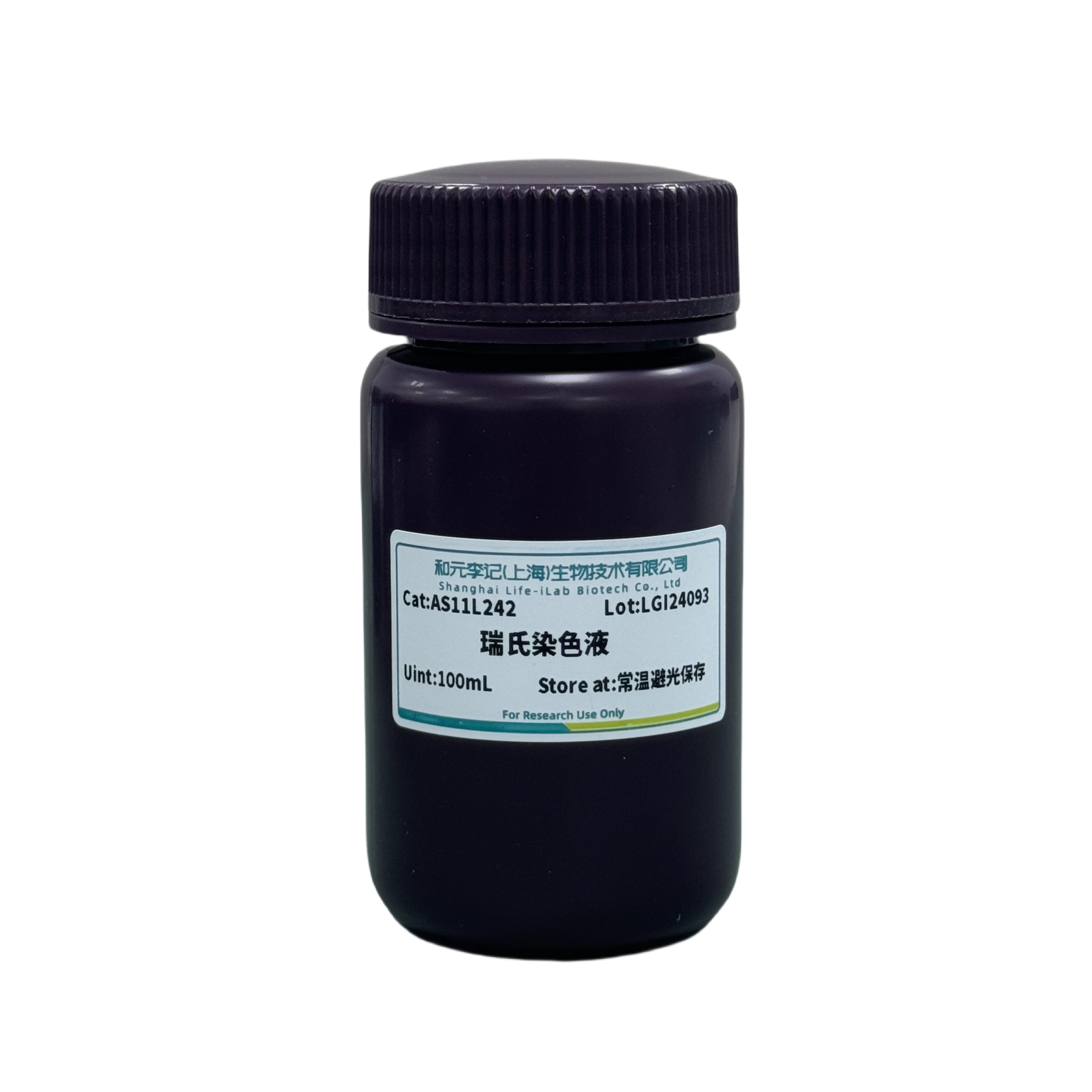
product description
Rui's dye is composed of alkaline dye methylene blue and acidic dye eosin, and different cellular components have varying affinities for the dye. Its characteristics are: hemoglobin and eosinophilic granules are alkaline proteins that bind with the acidic dye eosin and stain pink; The cytoplasm of nuclear proteins, lymphocytes, and eosinophils is acidic and binds to alkaline dyes such as methylene blue or azurol, dyeing them purple blue or blue; Neutral particles are in an isoelectric state and can bind to both eosin and methylene blue, resulting in a light purple red color.
Protein is one of the most important components in cells, with amphoteric electrolyte properties, and its charge varies with the pH value of the solution. In a slightly acidic environment, the positive charge increases, making it easy to bind with eosin. Red blood cells and eosinophils stain reddish, and the nucleus appears light blue or unstained; In a slightly alkaline environment, negative charges increase and easily combine with methylene blue. All cells appear gray blue with dark particles, while eosinophilic particles appear dark brown or even brownish black. Neutral particles are thicker and appear purple black. Therefore, the diluted dye solution must be diluted with phosphate buffer solution (pH 6.4-6.8), and the rinsing water should be near neutral, otherwise it may cause abnormal staining of cells and difficult recognition of morphology.
ordering information
product name | Item number | specifications |
Wright stain | AS11L242 | 100mL |
Transportation and storage
Transport at room temperature. Store at room temperature and avoid light, with a shelf life of 12 months.
Usage
1. Smear and fix: Spread the cells evenly on a clean glass slide, and fix them after drying.
The choice of fixative depends on the specific situation, and most cells can be fixed with methanol. Methanol fixation: After drying the smear, immerse it in methanol for 15-30 minutes for fixation.
2. Staining:
(1) After fixation, ventilate and dry at room temperature. Place the slide in the dye vat and prepare for dyeing.
(2) After dropping the Wright staining solution onto the glass slide, let it evenly cover the cells on the slide. The specific staining time depends on the cells, usually 2-3 minutes, and it is best to observe under a microscope during staining. When the Wright staining solution shows a tendency to turn red, quickly add an equal amount of phosphate buffer solution (pH 6.4-6.8) to the Wright staining solution and continue staining for 2-3 minutes.
(3) After staining, rinse the slide with phosphate buffer solution (pH 6.4-6.8) first, and then rinse with a small stream of water to prevent a large number of cells from falling off the slide and affecting subsequent observation. After the film processing is completed, place the slide in a cool place to air dry.
3. Sealing: Neutral gum sealing, made into a permanent smear.
4. Microscopic observation: Place the dried slide under a microscope for observation.
Staining result: The nucleus is stained blue, while the cytoplasm is stained red.
matters needing attention
1. This product is only for scientific experimental research and should not be used in clinical diagnosis, treatment, or other fields.
2. For your safety and health, please wear lab clothes and disposable gloves when operating.
3. Fix the blood smear after it has dried completely, otherwise the cells are prone to shedding during the staining process.
4. When rinsing, rinse with running water and do not pour out the dye solution first to prevent the dye from depositing on the blood smear. The rinsing time should not be too long to prevent discoloration. If dye particles are deposited on the blood smear, methanol can be added dropwise and immediately rinsed with running water.
5. If the dyeing is too light, it can be re dyed. When re dyeing, buffer solution should be added first, followed by dye solution. Deep staining can be washed or soaked with running water, or decolorized with methanol.
6. It is recommended to take 1-2 samples for preliminary experiments when using this kit for the first time.
Related product recommendations
Universal tissue cell fixative (4% paraformaldehyde) (item number: AC28L112)
Anti fluorescence quencher (item number: AC28L512)
Ruishi Jimsa staining solution (item number: AS11L212)
Jimsa staining solution (item number: AS11L232)
This product is only for scientific experimental research and should not be used in clinical diagnosis, treatment, or other fields.


 Back
Back
 Back
Back


