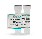
"Heyuan Liji" is a joint venture between Heyuan Biotechnology (stock code: 688238) and Liji Biotechnology, specializing in the "Liji Biotechnology" and "Life-ilab" reagent brands

Language: English

HOT PRODUCTS

RECOMMENDATION Cell Biology Common medium Transfection reagent Protein and immunity Nucleic acid electrophoresis

TECHNICAL SERVICE

Services Vector for gene therapy Genetic manipulation Cell experiments Animal testing Other special services

转录组学

原文链接:和元生物——转录组学转录组测序(RNA-Seq)是指利用第二代高通量测序技术进行cDNA测序,全面快速地获取某一物种特定器官或组织在某一状态下的几乎所有转录本。 随着后基因组时代的到来, 转录组学、蛋白质组学、代谢组学 等各种组学技术相继出现,其中转录组学是率先发展起来以及应用最广泛的技术。RNA-Seq具有以下优势:(1)通量高,运用第二代测序平台可得到几个到几百亿个碱基序列,可以达到覆盖整个基因组或转录组的要求;(2)灵敏度高,可以检测细胞中少至几个拷贝的稀有转录本;(3)分辨率高,RNA一Seq的分辨率能达到单个碱基,准确度好,同时不存在传统微阵列杂交的荧光模拟信号带来的交叉反应和背景噪音问题;(4)不受限制性,可以对任意物种进行全转录组分析,无需预先设计特异性探针,能够直接对任何物种进行转录组分析。同时能够检测未知基因,发现新的转录本,并准确地识别可变剪切位点及SNP、UTR区域。RNA-seq技术能够在单核酸水平对特定物种的整体转录活动进行检测,从而全面快速地获得该物种在某一状态下的几乎所有转录本信息。由于转录组测序可以得到全部RNA转录本的丰度信息,加之准确度又高,使得它具有十分广泛的应用领域。主要应用于:(1)检测新的转录本,包括未知转录本和稀有转录本;(2)基因转录水平研究,如基因表达量、不同样本间差异表达;(3)非编码区域功能研究,如microRNA、非编码长RNA (IncRNA)、RNA编辑;(4)转录本结构变异研究,如可变剪接、基因融合;(5)开发SNPs和SSR等。

Details

miRNA测序

原文链接:和元生物——miRNA测序小RNA是生物体内一类具有重要调控功能的非编码短小RNA的总称。大量研究已经证实,小RNA几乎参与调控了动植物所有的生命过程,包括细胞增殖,分化,凋亡等,并且与人类疾病的发生发展密切相关。小RNA测序正是对这一类重要的调控小RNA——特别是microRNA(miRNA)展开分析,利用高通量测序技术对样本中18~30nt的小RNA序列进行鉴定和分析。技术优势文库优化:针对动植物miRNA序列的不同特别,优化文库制备流程,最大程度地富集样本小RNA序列独家分析软件:拥有自主开发的数据分析软件ACGT101-miR,可靠性已被数百个实验项目检验;生成图表可直接用于论文撰写。项目经验丰富:国内最早提供小RNA测序服务的公司之一,用户已发表上百篇小RNA测序相关论文。技术路线Total RNA3’及5’接头连接反转录PCR扩增PAGE纯化上机测序(HiSeq 2500,SE50)数据分析分析内容1、数据质控:测序质量值分布统计;测序碱基质量控制;测序数据产出统计。2. 可比对测序数据的获得3. miRNA数据库以及测序物种基因组比对4. 与其他RNA数据库的比对5. 预测全新的miRNA6. miRNA序列本身相关分析7. miRNA定量分析,多样品miRNA的表达差异分析;差异表达miRNA筛选;差异表达miRNA统计;miRNA表达模式聚类分析(仅限多样本项目)。8. 重复相关性检测(仅限生物学重复样本)。9. 差异表达miRNA的靶基因预测(植物采用Targetfinder软件,动物采用Targetscan和miRanda软件0)10. 差异表达miRNA的靶基因GO,KEGG注释与GO,KEGG富集性以及pathway 通路分析和pathway network分析。(仅限多样本项目)差异表达miRNA的靶基因GO,KEGG注释与GO,KEGG富集性以及pathway 通路分析和pathway network分析。(仅限多样本项目)样本类型细胞,组织,体液、血清、血浆、全血,总RNA等建议总RNA起始量:5 μg,最低2.5 μg,浓度≥120 ng/μL样本类型1.Wu S, Li Y, Chen S, Liang S, Ren X, Guo W, Sun Q, Yang X. (2017) Effect of dietary Astragalus Polysaccharide supplements on testicular piRNA expression profiles of breeding cocks. International Journal of Biological Macromolecules 103(1), 957-964.2.Tritten L, Tam M, Vargas M, Jardim A, Stevenson MM, Keiser J, Geary TG. (2017) Excretory/secretory products from the gastrointestinal nematode Trichuris muris. Experimental Parasitology 178(1), 30-36.3.Sun J, Yao L, Chen T, Xi Q, Zhang Y. (2017) The effect of dietary ginseng polysaccharide supplementation on the immune responses involved in porcine milk-derived esRNAs. bioRxiv [Epub ahead of print].4.Ghorecha V, Zheng Y, Liu L, Sunkar R, Krishnayya NSR. (2017) MicroRNA dynamics in a wild and cultivated species of Convolvulaceae exposed to drought stress. Physiology and Molecular Biology of Plants 23(2), 291-300.5.Li H, Peng T, Wang Q, Wu Y, Chang J, Zhang M, Tang G, Li C. (2017) Development of Incompletely Fused Carpels in Maize Ovary Revealed by miRNA, Target Gene and Phytohormone Analysis. Frontiers in Plant Science 8(1), 463.案例展示同源四倍体和二倍体水稻花粉发育过程中的小RNA分析与比较1.研究背景MicroRNAs (miRNAs)通过抑制其靶基因来调控植物基因的表达,且在植物生殖中起着重要的作用。然而,目前关于同源四倍体水稻miRNA组分析的研究是相当有限的。2.研究结果在这项研究中,研究人员运用小RNA测序对二倍体和多倍体水稻花粉发育过程中的miRNA组进行分析。研究结果表明,与二倍体水稻相比,四倍体水稻中共检测出172种差异表达的miRNAs(DEM),以及57种miRNA在同源四倍体水稻中特异性表达。在这172种DEM中,115种miRNA上调,61种miRNA下调。上调DEM的靶基因GO分析表明,它们的功能富集在减数分裂前间期的膜运输、减数分裂繁殖和单小孢子阶段的核苷酸结合。 osa-miR5788和osa-miR1432-5p_R+1在减数分裂时上调,它们的靶基因揭示了减数分裂相关基因的相互作用,也暗示了它们可能参与染色体行为相关的基因调控。此外,在同源四倍体水稻的花粉发育过程中,发现了丰富的与转座因子相关的24 nt 的siRNA;但是,它们在二倍体水稻中的含量明显下降,表明24 nt的siRNA在花粉发育中可能发挥作用。 图 花粉发育过程中不同阶段miRNA表达的Venn图分析本研究的结果为miRNA在同源四倍体水稻花粉发育过程中所起的作用,以及其与花粉不育性之间的关系提供了新的见解,为理解小RNA表达谱对多倍体的影响奠定了基础。参考文献Li, et al. (2016) Comparative Small RNA Analysis of Pollen Development in Autotetraploid and Diploid Rice. InternationalJournal of Molecular Sciences. 17, 499; doi:10.3390/ijms17040499结果展示1.差异miRNA上下调统计分析差异基因上下调频数统计用于判断不同实验条件下差异表达miRNA的个数。其中横坐标表示比较组信息,纵坐标表示上下调miRNA的数目,红色代表上调miRNA,蓝色代表下调miRNA,其中数字代表上下调miRNA的数目。 2.差异miRNA聚类分析差异miRNA聚类分析用于判断miRNA在不同实验条件下调控的聚类模式。根据样品miRNA表达谱的相近程度,将miRNA进行聚类分析,直观地展示miRNA在不同样品(或是不同处理)中的表达情况,由此获取生物学相关信息。不同的颜色表示不同的miRNA表达水平,颜色由蓝色经由白色至红色表示表达量从低到高。红色表示高表达基因,深蓝色表示低表达基因。 3.差异miRNA韦恩图通过不同比较组之间差异基因韦恩图可以直观地显示出不同比较组之间共同的和特有的差异表达miRNA的个数,差异基因韦恩图具有明显的生物学意义,比如是相同对照不同处理的实验设计情况,可以将不同处理下的差异基因进行比较。4.GO富集性柱状图miRNA靶基因的GO富集柱状图:用于反映在生物过程(biological process)、细胞组分(cellular component)和分子功能(molecular function)富集的GO term上差异基因的个数分布情况。5.GO富集性散点图对差异miRNA进行GO富集分析并以散点图展示,Rich factor表示位于该GO的差异基因个数/位于该GO的总基因数,P值越小,GO富集程度越高。常见问题 1.miRNA命名规则是怎样的?关于miRNA命名,是根据miRBase的命名规则:物种拉丁名3字母缩写-miR/MIR-编号(植物),miR表示的是microRNA成熟体,植物的前体使用MIR。参见miRBase官网:目前已经不再使用*来标记microRNA与其发夹前体互补配对位置的互补序列,而是使用“-3p”与“-5p”作为区分这两条序列的后缀替代旧的的命名法。具体参考17.0版本出来时,miRbase数据库的blog文章:http://www.mirbase.org/blog/2011/04/mirbase-17-released/ 2.如何筛选差异基因?可以在筛选的时候可能需要参考该参数,一般如果专注于发现,那么全部保留,如果专注于差异表达,可以仅保留high和middle拷贝的。log2(Treat/Control)>0表示上调,1表示上调2倍,0表示上调,1表示上调2倍,3.ACGT101程序的优势有哪些?优势有以下几点:1)分析全程兼顾测序质量打分(phred score);2)比对已报道成熟体之前先与已报道前体序列比对,除了发现全新的miRNA序列,还可以发现另一末端的成熟体序列,以及已报道成熟体的侧翼miRNA序列;3)与其他RNA数据库的比对过滤在于miRBase数据库比对之后,避免了滤除那些少量比对上编码区域的已报道miRNA;4)Table 5结果数据信息丰富全面,全部结果都有迹可循;5)100篇客户参考文献。

Details

lncRNA测序

原文链接:和元生物——lncRNA测序简介 长链非编码RNA(lncRNA)是一类长度大于200 nt 的非编码RNA(ncRNA),广泛存在于各种生物体内,在表观遗传调控、细胞周期调控和细胞分化调控等众多生命活动中发挥重要作用,与动植物的生长发育,人类的疾病发生有着密切关系,也可作为疾病诊断的标志物或是重要靶点。利用高通量测序技术进行lncRNA 测序及生物信息学分析,可快速准确地发现那些具有重要调控功能的lncRNA,分析其与特定生物学过程的关系,深入探索lncRNA的功能及其表达调控机制。包括竞争结合miRNA的ceRNA调控机制,及对染色质结构进行一系列调控如增强子、启动子、绝缘子等。 样本起始量与送样建议样本类型起始量动物及临床脏器组织/脑组织等>20mg动物及临床皮肤/骨/血管/脂肪组织等>100mg植物叶片组织/花>200mg植物根/茎/果实/种子>500mg原代细胞/细胞系>5×106个中性粒细胞/嗜酸性粒细胞/嗜碱性粒细胞>5×107个外泌体样本>1×108个血清/血浆/脑脊液/关节积液/卵泡液>2mL细胞培养上清液>20mL尿液>30mL总RNA>1μg且RIN>7.0注意事项:① 组织样本建议保存在RNAlater、RNAHold、RNAProtect等相关组织保存液中,然后-80℃保存或干冰寄送;② 细胞样本使用TRIzol等裂解液充分裂解之后,-80℃保存或干冰寄送③ 更加详细的样本准备指南,请联系在线客服 生物信息学分析流程与分析内容lncRNA分析内容 备注 测序数据质控测序原始数据量及cleandata数据量统计测序质量Q20 Q30 GC含量统计,去除原始下机数据中的接头、低质量及污染序列基因组比对参考基因组比对/区域比对染色体密度分布表达分析基因/转录本表达总谱样本基因表达值分布统计样本不同基因表达值区间分布统计样本基因转录本覆盖深度统计基因表达值密度图基因表达水平盒型图样本相关性分析Pearson/Sperman相关性分析PCA聚类分析mRNA分析基因差异分析转录本差异分析差异基因火山图差异基因整体统计图差异基因聚类热图差异基因GO富集分析差异基因KEGG富集分析GSEA分析lncRNA分析转录本重构和lncRNA预测lncRNA表达分析lncRNA差异表达分析差异lncRNA火山图差异lncRNA整体统计差异lncRNA聚类热图lncRNA和mRNA结构特征比较lncRNA和mRNA长度比较lncRNA和mRNA的ORF分布lncRNA和mRNA外显子数目统计lncRNA和mRNA表达水平统计lncRNA靶基因预测lncRNA靶向差异基因GO富集分析lncRNA靶向差异基因KEGG富集分析SNP/INDEL分析SNP/INDEL数目统计SNP/INDEL所在位置SNP突变型态统计基因区域SNP和INDEL注释可变剪切分析可变剪切类型统计可变剪切可视化 应用场景与案例 应用场景1:启动子/增强子调控适用范围:非线性转录调控、新顺式作用元件发现及功能鉴定、组织特异性启动子筛选及确定等研究lncRNA的自身转录会干扰其邻近编码蛋白的基因的转录。上游lncRNA转录时,会穿越邻近靶基因的启动子区,干扰了转录因子与靶基因的启动子结合,从而抑制靶基因的转录。此外,lncRNA具有促进增强子环化和激活基因表达的功能。在没有成环的情况下增强子处于非活性状态。应用场景2:与蛋白修饰/DNA甲基化/m6A联合适用范围:临床医学、基础医学、植物遗传育种等研究lncRNA参与表观调控,可能是招募一些染色质重构复合体来介导基因沉默,尤其是一些与组蛋白修饰相关的组蛋白甲基转移酶的调控。此外,lncRNA还能与一些DNA甲基化和去甲基化酶结合使得基因的启动子区发生甲基化的变化从而影响基因表达。应用场景3:ceRNA调控机制适用范围:环境应激、临床医学、植物遗传育种、疾病早期诊断等研究ceRNA全称competing endogenous RNA,是一种能够竞争结合RNA的作用元件。通常lncRNA和circRNA会竞争结合miRNA,我们一般把lncRNA和circRNA可以称作ceRNA。ceRNA调控网络全称ceRNAregulation network,指的是有ceRNA参与的整个调控网络cascade。而ceRNA分析指的是对整个ceRNA调控网络进行分析。一般有circRNA-miRNA-mRNA分析或lncRNA-miRNA-mRNA分析。 项目流程 和元生物真核有参转录组测序可以为研究人员提供从样本提取、建库测序、数据分析等一系列完整的服务流程,提供高质量的数据结果,并为后续研究提供强有力的参考依据。

Details

单细胞测序

原文链接:和元生物——单细胞测序单细胞测序技术,是在单个细胞水平上对基因组、转录组、表观组进行高通量测序分析的一项新技术。它能够揭示单个细胞的基因结构和基因表达状态,反映细胞间的异质性,在肿瘤、发育生物学、微生物学、神经科学等领域发挥重要作用,正成为生命科学研究的焦点。单细胞测序工作流程单细胞测序的工作流程包括 4 个关键步骤 :1) 单细胞制备,2)单细胞分离和文库制备,3)测序和初级分析,4)数据可视化与解读。在整个工作流程中,有一些影响结果并决定研究能否成功的实验注意事项和关键步骤。想要获得准确的数据,得出有意义的结论,需要精心设计实验,并按要求开展实验。在过去的十年中,随着细胞分离新技测序新方法术和单细胞和新应用的发展,单细胞鉴定和研究领域取得了重大进展。随着单细胞分离和检测的选项越来越多,实验方案的多样性显著增加,每种方案都有其固有的优缺点。因此,研究人员面临着诸多决策问题,例如细胞通量、测序深度、所需转录本长度、是否应纳入表观遗传或蛋白水平测量等。想要充分利用单细胞测序来阐明复杂的生物系统,精心设计实验和优化工作流程的每个步骤至关重要。研究人员必须有明确的生物学目标和合理的实验设计,才能选择出针对其研究问题的最佳方法。

Details

Eukaryotic transcriptome sequencing

Original link:Metaorganisms - Eukaryotic Transcriptome Sequencingbrief introductionTranscriptome broadly refers to all transcripts of a specific cell in a certain functional state, including mRNA and non coding RNA (ncRNA). It is an inevitable link between genomic genetic information and biological functions. Transcriptome sequencing in eukaryotes is based on high-throughput sequencing, which can quickly obtain a collection of all transcripts of a specific cell or tissue in a certain state of a species, used for studying gene structure and function, variable splicing, and predicting new transcripts.ranscriptome research can study gene function and structure at the overall level, and has become a priority research method to reveal the mechanisms of biological growth and development regulation and adaptation to stress, biological evolution laws, important mechanisms of disease occurrence and development, and discover key targets of pathogenic gene regulation. Currently, it has been widely applied in various fields such as basic research, clinical diagnosis and drug development, animal and plant breeding, etc.characteristicAbility to handle complex samplesRich experience in library construction, incorporating library homogenization technologyAssist clients in quickly and accurately conducting bioinformatics analysisCan flexibly customize information analysis based on customer needsInitial sample size and sample delivery recommendationsSample typeinitiation massAnimal and clinical organ tissues/brain tissues, etc>20mgAnimal and clinical skin/bone/blood vessels/adipose tissue, etc>100mgPlant leaf tissue/flowers>200mgPlant roots/stems/fruits/seeds>500mgPrimary cells/cell lines>5 x 10⁶ piecesNeutrophils/eosinophils/basophils>5 x 107 piecesTotal RNA>1 μ g and RIN>7.0matters needing attention:① It is recommended to store the tissue samples in relevant tissue preservation solutions such as RNAlate, RNAOld, RNAProtect, etc., and then store them at -80 ℃ or send them on dry ice;② After sufficient lysis using TRIzol or other lysis solutions, the cell samples are stored at -80 ℃ or sent on dry ice③ For more detailed sample preparation guidelines, please contact online customer serviceBioinformatics analysis process and contentReferenced transcriptomeAnalysis contentremarksSequencing data quality controlRemove connector sequences, contaminated sequences, and low-quality error sequences from the original offline dataData volume statistics and quality evaluationSequence alignment and transcript reconstructionReference genome alignmentCompare the proportion of offline data to the upper genomeDistribution of reference genome alignment regionsStatistical comparison of the proportion of exons and introns in the genome sequenceReference sequence chromosome density distributionStatistical distribution of alignment sequences on chromosomesTranscriptome reconstructionContains sequence merged. fa and. gtf filesGene/transcript overall expression analysisGene expression summary tableTranscript expression summary tableBox plot of gene and transcript expression distributionStatistical distribution of gene and transcript expression intervalsTranscript coverage depth statisticsDistribution density map of gene and transcript expression levelsDifferential expression gene/transcriptome analysis (sample size ≥ 2)Differential expression genes and transcripts statistical bar chartDifferential expression genes and transcriptome expression profilesDifferential expression genes and transcriptome volcano mapCluster heatmap of differentially expressed genes and transcriptsGO enrichment analysis of differentially expressed genesIncluding GO enrichment bar charts, scatter plots, radar charts, etcEnrichment analysis of differentially expressed genes KEGGIncluding KEGG pathway enrichment scatter plots, pathway maps, radar maps, etcEnrichment analysis of differentially expressed genes Reactome (including only 19 common species)Including Reactome enrichment scatter plots, bar charts, etcEnrichment analysis of differentially expressed genes DO (disease annotation database) (including only human species)Including scatter plots, bar charts, etc. for DO database enrichmentstructural analysisVariable splicing analysisBy default, ASprofile variable shear analysis results are provided, and rMATS differential variable shear analysis results can be provided for free after salesSNP/InDel analysisSample correlation (sample size ≥ 2)Correlation coefficient chart and PCA (principal component analysis) chartApplication scenarios and casesApplication Scenario 1:Differential gene screening and functional analysisApplicable scope:Any direction including clinical medicine, basic medicine, biochemistry, animal and plant and fungal researchBy using eukaryotic transcriptome sequencing, differentially expressed genes can be screened by comparing the gene expression levels between the experimental group and the control group. Then, further locking of differentially expressed genes is carried out, such as GO, KEGG enrichment analysis and GSEA analysis, combined with published literature in Pubmed and some star molecules accumulated in the research group to annotate the functions of differentially expressed genes, and further analyze the functional genes of interest. After entering the experimental validation stage, qPCR, Northern, Western Blot, FISH validation, gene knockout, and overexpression can be performed on the screened differentially expressed genes.Application Scenario 2:Time series analysis or concentration gradient analysisApplicable scope:Clinical samples, cell samples, animal and plant samples with multiple time periods, or samples treated with different drug concentrationsIn the process of transcriptome data analysis, there is a special type of experimental design. Collect experimental samples from different time periods or test samples with different concentration gradients of drugs, reagents, etc. Subsequently, studying the expression patterns of different genes at different time periods or concentration gradients is commonly referred to as "time series analysis"Application Scenario 3:Discovery of upstream regulatory genes such as transcription factors/regulatory factors/splicing factorsApplicable scope:Any research direction including clinical medicine, basic medicine, biochemistry, animal and plant research, etcConventional transcriptome differential analysis is highly likely to yield a large number of differentially expressed genes, which poses a challenge for target localization in later experimental validation. Transcription factors are a great entry point without specific pathways of interest or star molecules. Transcription factors can regulate genomic DNA openness, recruit RNA polymerase for transcription processes, recruit cofactors to regulate specific transcription stages, and regulate various life processes such as immune response and developmental patterns. Therefore, analyzing the expression and regulatory activity of transcription factors is of great significance for deciphering complex life activities. Other regulatory factors, including variable splicing and other regulatory genes, can also participate in upstream regulation.Application scenario 4: Large sample researchApplicable scope:Animal and plant breeding, genetic populations and species origins, population cohorts and biomarker miningWith the rapid development of sequencing technology, transcriptome sequencing studies with a small number of samples are no longer able to explain complex biological problems. Researchers have begun to use large sample sizes of transcriptome samples, combined with statistical and machine learning methods, to identify core genes that conform to specific patterns and research objectives. Using methods such as Mendelian randomization, correlation analysis, linear regression, LASSO regression, Cox regression, etc., to analyze gene or genomic diversity in different samples and explore deeper and more comprehensive biological significance.Project ProcessTranscriptome sequencing with eukaryotic involvement in metaorganisms can provide researchers with a complete range of service processes, including sample extraction, library sequencing, and data analysis, providing high-quality data results and providing strong reference for subsequent researchAt the same time, the cloud analysis of metabiotic transcriptome has been fully upgraded, providing customers with a variety of analysis content to meet the standard and personalized analysis needs of researchers.

Details

CircRNA sequencing

Original link:Metaorganisms - circRNA sequencingbrief introductionCovalent closed, single stranded circular RNA (circRNA) is a relatively special type of RNA that does not have a free 5 'cap structure or a 3' poly (A) structure, and is not sensitive to nucleases, making it more stable than regular linear RNA. With the rapid development of high-throughput sequencing and bioinformatics analysis technology, thousands of circRNAs have been identified in different species, most of which are conserved and stable between different species, and the expression of circRNAs is cell specific, tissue specific, and developmental stage specific. In addition to the conventional ceRNA regulatory mechanism that competes for miRNA binding, circRNA also has new functions such as translation peptides, which have great potential in medical and animal and plant basic research and later transformation.Initial sample size and sample delivery recommendationsSample typeinitiation massAnimal and clinical organ tissues/brain tissues, etc>20mgAnimal and clinical skin/bone/blood vessels/adipose tissue, etc>100mgPlant leaf tissue/flowers>200mgPlant roots/stems/fruits/seeds>500mgPrimary cells/cell lines>5 x 10⁶ piecesNeutrophils/eosinophils/basophils>5 x 107 piecesExtracellular vesicle samples>1x10⁸ piecesSerum/plasma/cerebrospinal fluid/joint effusion/follicular fluid>2mLcell culture supernatant >20mLurine>30mLTotal RNA>1 μ g and RIN>7.0matters needing attention:① It is recommended to store the tissue samples in relevant tissue preservation solutions such as RNAlate, RNAOld, RNAProtect, etc., and then store them at -80 ℃ or send them on dry ice;② After sufficient lysis using TRIzol or other lysis solutions, the cell samples are stored at -80 ℃ or sent on dry ice;③ For more detailed sample preparation guidelines, please contact online customer serviceBioinformatics analysis process and contentcircRNAAnalysis content remarks Sequencing data quality controlRaw data quality control statistical tableReference genome alignment analysisReference genome gene statistics tableReference genome alignment reads statistics tableStatistical Table of the Number of CircRNA Identification SamplesStatistical chart of reference genome alignment region distributionReference genome nonlinear alignment region distribution statistical chartQuantitative analysis of circRNA identificationCircRNA fitting sequenceCircRNA expression profileBSJ statistical table for circRNACircRNA statistical analysisCircRNA type statistical pie chartStatistical boxplot of circRNA expression distributionDistribution map of circRNA expression densityStatistical chart of the distribution of circRNA chromosome numbersStatistical chart of the number of circRNA exonsStatistical chart of the number of circRNAs in parental genesCircRNA differential analysisStatistical chart of the number of differential circRNAsAnalysis of differential expression of circRNA in CaseVSControl comparison groupCase VSControl comparison group circRNA volcano mapCase VSControl comparison group circRNA heatmapCaseVSControl comparison group circRNA scatter plotApplication scenarios and casesApplication scenario 1: Structural molecule recruitment of core proteins and nucleic acidsApplicable scope: Any research direction such as basic medicine, biochemistry, and molecular biologyLncRNA and circRNA sometimes act as special RNA molecules, forming protein protein complexes or backbone molecules of protein nucleic acids, exerting a series of regulatory effects on downstream molecules. Some circRNAs recruit ubiquitinases to degrade downstream proteins, or some lncRNAs recruit transcription factors to bind to the promoter region of the target gene to activate downstream gene expression.Application scenario 2: ExosomesScope of application: Any research direction in clinical and translational medicine, basic medicine, etcDue to the fragmented form of lncRNA in exosomes, this random fragment is not only difficult to detect, but also has lower concentrations and poor repeatability, making lncRNA not one of the hot molecules in exosome research. However, due to its stability, circRNA can not only be isolated from extracellular vesicles in body fluid samples as a disease marker, but also be used for later treatment of extracellular vesicles, making it one of the more popular sub research directions for circRNA in the post ceRNA era. One direction is disease biomarkers, which are generally studied in large sample cohorts. One is the research direction of late stage transformation and mechanism, including the molecular mechanism of extracellular vesicle receptor cells and disease treatment.Application scenario 3: Translating peptidesScope of application: Any research direction including clinical and translational medicine, basic medicine, biochemistry and molecular biology, etcThere is an untranslated RNA sequence on circRNA, called IRES, which can fold into a structure similar to the starting tRNA and recruit more ribosomes. Under normal circumstances, IRES does not bind to eIF, but rather to a class of proteins called ITAF. The function of ITAF is to recruit ribosomes into the internal structure of circRNA to initiate protein translation. In addition, there may also be IRES enhancers, similar to UTR elements of circ-ZNF609. Some studies have also found through transcriptome sequencing and transcriptome sequencing that non coding RNA LINC-PINT can be translated into functional peptides after forming circular RNA.Application scenario 4: ceRNA regulatory mechanismThe full name of ceRNA is competitive endogenous RNA, which is an element that can compete for binding to RNA. Usually, lncRNA and circRNA compete to bind to miRNA, and we generally refer to lncRNA and circRNA as ceRNA. The full name of ceRNA regulation network is ceRNA regulation network, which refers to the entire regulatory network cascade in which ceRNA participates. And ceRNA analysis refers to the analysis of the entire ceRNA regulatory network. Generally, there are circRNA miRNA mRNA analysis or lncRNA miRNA mRNA analysis.Project Process Project ProcessTranscriptome sequencing with eukaryotic involvement in metaorganisms can provide researchers with a complete range of service processes, including sample extraction, library sequencing, and data analysis, providing high-quality data results and providing strong reference for subsequent research.

Details

Lentiviral vector

原文链接:Metaorganisms - lentiviral vectorsLentivirus is a type of viral vector modified from human immunodeficiency virus (HIV), which is a type of retrovirus. The genome is RNA, and its toxic genes have been removed and replaced by exogenous target genes. Lentivirus belongs to the pseudotype virus, which can integrate foreign genes into the genome to achieve stable expression and has the characteristics of infecting dividing and non dividing cells.After the lentivirus genome enters the cell, it is reverse transcribed into DNA in the cytoplasm, forming a DNA pre integration complex. After entering the nucleus, DNA is integrated into the cell genome. The integrated DNA is transcribed into mRNA and returned to the cytoplasm to express the target protein or produce small RNA. The gene expression or small RNA interference mediated by lentivirus is sustained and stable, and divides with the division of the cell genome (Figure 1). Figure 1 Process of Slow Virus Packaging and Infection of Cells  Slow virus vectors have the characteristics of wide host range, low immunogenicity, large gene capacity, and long-term expression. Capable of effectively infecting various types of cells in culture, including tumor cells, liver cells, myocardial cells, neurons, endothelial cells, stem cells, etc.  The infection of lentivirus has the characteristic of integration, which can effectively integrate foreign genes into the host chromosome, achieve persistent expression, and thus construct stable cell lines for the study of gene cellular function. The use of lentiviral vectors to modify T cells can be used for CAR-T cell therapy.Slow virus packaging and purification  Slow virus packaging uses a three plasmid or four plasmid system for packaging, collects cell culture supernatant, and concentrates, purifies, and collects viruses through high-speed centrifugation.Slow virus titer detection  Detection method: Quantitative PCR is used to detect the copy number of exogenous DNA in the cell genome after infection  Experimental principle: lentivirus mediates the integration of exogenous genes into the target cell genome through reverse transcriptionAdvantages of lentiviral vectors① Long expression time: Slow viruses can integrate exogenous genes into the host cell genome, achieving stable gene expression for a long time without loss during cell division and passage. It is the preferred choice for cell experiments and is commonly used to construct stable strains.② High safety: No pathogenicity was found, and lentiviral vectors were used to modify T cells for CAR-T cell therapy.③ Low immunogenicity: Direct injection into live tissue does not easily cause an immune response and is suitable for animal experiments.Comparison of Biological Characteristics of Different VirusesCarrier selection  HarmonyBio has a rich range of lentiviral products for manipulating both coding and non coding genes, such as lncRNA microRNA、circRNA, The following slow virus vectors are available for you to choose from (not limited to this list):Regulatory methodsCarrier numberCarrier name (order of carrier components)Eukaryotic resistancefluorescencePromoterDefault labelCarrier capacityOverexpressionGL127pSLenti-CMV-EGFP-3xFLAG-WPREN/AEGFPCMV3FLAG4.8kbGL121pSLenti-EF1-EGFP-CMV-MCS-WPREN/AEGFPCMVN/A4.4kbGL129pSLenti-CMV-mCherry-3xFLAG-WPREN/AmCherryCMV3FLAG4.8kbGL119pSLenti-CMV-MCS-3xFLAG-PGK-Puro-WPREPuroN/ACMV3FLAG4.4kbGL120pSLenti-SFH-EGFP-P2A-Puro-CMV-MCS-3xFLAG-WPREPuroEGFPCMV3FLAG3.5kbGL107pSLenti-EF1-EGFP-P2A-Puro-CMV-MCS-3xFLAG-WPREPuroEGFPCMV3FLAG3.7kbGL109pSLenti-EF1-EGFP-F2A-Puro-CMV-MCS-WPREPuroEGFPCMVN/A3.8kbGL122pSLenti-EF1-EGFP-F2A-BSR-CMV-MCS-WPREblasticidinEGFPCMVN/A3.9kbGL130pSLenti-CMV-EGFP-3xFLAG-PGK-Puro-WPREPuroEGFPCMV3FLAG3.7kbGL132pSLenti-EF1-EGFP-F2A-Puro-WPRE2-CMV-MCSPuroEGFPCMVN/A3.7kbGL123pSLenti-EF1-mCherry-P2A-Puro-CMV-MCS-3xFLAG-WPREPuromCherryCMV3FLAG3.7kbGL125pSLenti-CMV-mCherry-3xFLAG-PGK-Puro-WPREPuromCherryCMV3FLAG3.7kbGL133pSLenti-EF1-mCherry-F2A-Puro-WPRE2-CMV-MCSPuromCherryCMVN/A3.7kbH114pLenti-CMV-Luc2-IRES-Puro-WPREPuroN/ACMVN/A2.2kbGL124pSLenti-EF1-Luc2-F2A-Puro-CMV-MCS-WPREPuroN/ACMVN/A2.8kbExpressing creH126pLenti-CMV-NLS-Cre-3xFLAG-WPREN/AN/ACMV3FLAG3.9kbH15108pLenti-CMV-DIO-MCS-WPREN/AN/ACMV3FLAG5.3kbThree standardsH7656pLenti-CBh-3xFLAG-Luc2-tCMV-mNeonGreen-F2A-Puro-WPREPuromNeonGreenCBh3FLAG1.3kbH7657pLenti-CBh-3xFLAG-Luc2-tCMV-tdTomato-F2A-Puro-WPREPurotdTomatoCBh3FLAG0.6kbH9911pLenti-CBh-3xFLAG-Luc2-tCMV-mNeonGreen-F2A-BSR-WPREblasticidinmNeonGreenCBh3FLAG1.5kbH9912pLenti-CBh-3xFLAG-Luc2-tCMV-tdTomato-F2A-BSR-WPREblasticidintdTomatoCBh3FLAG0.8kbCircRNA overexpressionH8384pLenti-EF1-EGFP-F2A-Puro-CMV-S-circRNA-WPREN/AEGFPCMVN/A3.0kbH8807pLenti-CMV-S-circRNA-WPREN/AN/ACMVN/A4.8kbH8399pLenti-EF1-EGFP-F2A-Puro-CMV-L-circRNA-WPREPuroEGFPCMVN/A1.0kbH8810pLenti-CMV-L-circRNA-WPREN/AN/ACMVN/A2.8kbMiRNA overexpressionGL109pSLenti-EF1-EGFP-F2A-Puro-CMV-MCS-WPREPuroEGFPCMVN/AN/AH3919pCLenti-U6-miR30(miRNA)-CMV-EGFP-F2A-Puro-WPREPuroEGFPU6N/AN/AH119pLenti-CMV-TurboGFP-IRES-Puro-miR30(miRNA)-WPREPuroTurboGFPCMVN/AN/AH3928pCLenti-U6-miR30(miRNA)-CMV-mCherry-F2A-Puro-WPREPuromCherryU6N/AN/AH146pCLenti-EF1-Puro-CMV-EGFP-3xFLAG-Sponge(miRNA)-WPREPuroEGFPCMV3FLAGN/AH7505pCLenti-U6-TuD(miRNA)-CMV-EGFP-F2A-BSR-WPREblasticidinEGFPCMVN/AN/AH7506pCLenti-U6-TuD(miRNA)-CMV-EGFP-F2A-Puro-WPREPuroEGFPCMVN/AN/AOverexpression (Tet on)H125pLenti-TRE-EGFP-EF1-rtTA3-IRES-Puro-WPREPuroEGFPTREN/A2.0kbH121pLenti-TRE-EGFP-3xFLAG-PGK-Puro-WPREPuroEGFPTRE3FLAG3.3kbH2057pLenti-EF1-rtTA3-IRES-Puro-WPREPuroN/AEF1N/AN/AShRNA interferenceGL401pCLenti-U6-shRNA-CMV-Puro-WPREPuroN/AU6N/AN/AGL404pCLenti-U6-shRNA-CMV-EGFP-WPREN/AEGFPU6N/AN/AGL427pSLenti-U6-shRNA-CMV-EGFP-F2A-Puro-WPREPuroEGFPU6N/AN/AGL428pSLenti-U6-shRNA-CMV-mCherry-F2A-Puro-WPREPuromCherryU6N/AN/AH7615pCLenti-U6-shRNA-CMV-mCherry-F2A-BSR-WPREblasticidinmCherryU7N/AN/ACRISPR KnockoutH5070pLenti-U6-spgRNA v2.0-CMV-Puro-P2A-3xFLAG-spCas9-WPREPuroN/AU6N/AN/AH7072pLenti-U6-spgRNA v2.0-CMV-BSR-P2A-3xFLAG-spCas9-WPREblasticidinN/AU6N/AN/AH6825pLenti-U6-spgRNA v2.0-CMV-sfGFP-P2A-3xFLAG-spCas9-WPREN/AsfGFPU6N/AN/AH5068pLenti-U6-spgRNA v2.0-CMV-EGFP-WPREN/AEGFPU7N/AN/AH5450pLenti-CMV-Puro-P2A-3xFLAG-espCas9_1.1-WPREPuroN/ACMV3FLAGN/ACRISPRa transcriptional activationH9517pCLenti-U6-gRNA-MS2-EFS-dCas9-VP64-T2A-BSD-WPREblasticidinN/AU6N/AN/AE2577pCLenti-EF1a-MCP-P65-HSF1-T2A-Hygro-WPREhygromycinN/AEF1aN/AN/AH7281pLenti-CMV-dCas9-VP64-T2A-Puro-WPREPuroN/ACMVN/AN/AConventional applications  In vitro infected cells   Slow virus vectors can effectively infect various types of primary cells and most cell lines, including tumor cells, liver cells, myocardial cells, neurons, endothelial cells, stem cells, etc.   The infection of lentivirus has integration characteristics, which can effectively integrate foreign genes into the host chromosome, thus achieving persistent expression and constructing stable transgenic cell lines. In vitro cellular functional experiments, including cell proliferation, invasion, migration, apoptosis, etc., are conducted. Stable expression of target genes in tumor cell lines can further construct tumor animal models for in vivo gene function validation, pharmacokinetic analysis, in vivo imaging, and other detection to further study the occurrence, development, metastasis, and drug treatment effects of tumors in vivo. Common MOI ListCell nameChinese nameChronic viral infection (MOI)(Reference value)MGC80-3Human gastric cancer cells10HT-29human colon cancer cell20RKOHuman colon adenocarcinoma cells10Caki-1Human renal clear cell carcinoma skin metastatic cells105637Human bladder cancer cells10U-118 MGHuman astroblastoma10RWPE-1Normal human prostate epithelial cells20HeLaHuman cervical cancer cells10~20Ca SkiHuman cervical cancer intestinal metastatic cells10MCF7 [MCF-7]Human breast cancer cells10A549Human non-small cell lung cancer cells10NCI-H1299Human non-small cell lung cancer cells20U-87 MGHuman astroblastoma10U251Human glioma cells10A172Human glioblastoma cells10K-562Human chronic myeloid leukemia cells10HL-60Human myeloid leukemia cells10GES-1Human gastric epithelial cells20U266Human myeloma cells20CAL 27Human tongue squamous cell carcinoma cells20PC9Human lung cancer cells5PC14Human lung cancer cells10MADB-106Rat breast cancer cells20F98Rat glioma cells20MHCC-97HHuman liver cancer cells (high metastasis)10~20  MOI stands for multiplicity of infection, also known as the plural number of infections, which refers to the ratio of the number of viruses to cells during infection. Generally, in an experiment, when a certain cell is infected and reaches 80%, it is defined as the MOI value of that cell, that is, the ratio of the number of viruses to the number of cells; The amount of virus added (μ l)=number of cells x MOI/titer (.../ml) x 1000Application cases of lentivirus in tumor research1.Nature Communications. (IF=12.353). Shi Y,et.al. (2017). Tumour-associated macrophages secrete pleiotrophin to promote PTPRZ1 signalling in glioblastoma stem cells for tumour growth.[lentivirus, glioma]Slow virus interference: Lenti-shNT/shPTN/shPTPRZ12. Hepatology. (IF=14.079). Ma JZ,et.al. (2016). METTL14 suppresses the metastatic potential of HCC by modulating m6 A-dependent primary miRNA processing. [lentivirus, liver cancer]Slow virus overexpression/interference:pLV-CMV-PGK-EGFP-T2A-puro/ pLV-U6-shRNA-CMV-EGFP-T2A-puroThe application of lentivirus in the nervous system (in vivo injection)1.Nature Neuroscience. (IF=19.912). Ding XL,et.al. (2017). Activity-induced histone modifications govern Neurexin-1 mRNA splicing and memory preservation.[Slow virus, learning and memory]Virus type: lentivirusCarrier name:LV-Suv39h1-RNAi(Uncertain details)Injection site: DG area of hippocampusVirus injection volume:2 μL,9.98 × 108 TU/mLTesting time: 2 weeks2.Nature Communications. (IF=12.353). Yao XH,et.al. (2016). Electrical coupling regulates layer 1 interneuron microcircuit formation in the neocortex.[lentivirus, neural circuit]Virus type: lentivirusCarrier name:LV- CX36-shRNA-EGFP(Uncertain details)Injection site: P1 mouse neocortex L1Virus injection volume:1μLTesting time: 2 weeks3.Molecular Psychiatry. (IF=11.64). Guo DJ,et.al. (2019). Autism-like social deficit generated by Dock4 deficiency is rescued by restoration of Rac1 activity and NMDA receptor function. [Chronic virus, autism]Virus type: lentivirusCarrier name:Lenti-CMV-EGFP-P2A-3FLAG-Rac1Injection site: CA1 area of mouse hippocampusVirus injection volume: 1 μ LTesting time: 4 weeksSlow virus extension service The production and quality control of metabiotic lentiviruses have adopted internationally recognized standard processes, using a three plasmid or four plasmid system for packaging in 293T cells. The obtained virus particles were purified by ultracentrifugation and the viral gene copies were titrated by qPCR. In general, the titer of our lentivirus is between 108~109TU/ml, which can fully meet the requirements of various experiments.

Details

Adenovirus ADV

Original link:Harmony with Metaorganisms - Adenovirus ADVAdenovirus (ADV) is a linear double stranded DNA virus without an envelope, with a wide range of cell and tissue infectivity. The infection process is intense, making it suitable for experiments with high gene expression in a short period of time. Compared with other viruses, adenoviruses have the advantage of longer insertion fragments and stronger expression activity.  When used in vitro, adenovirus vectors have high transgenic efficiency (close to 100% transduction efficiency) and can transduce different types of cells, making them a good tool for delivering genes to a large number of difficult to transfect cells. However, when applied in vivo, due to the strong immunogenicity and severe infection of adenovirus, it often causes local tissue inflammation and immune response in animals, which affects the objectivity of animal signs and experimental results.ADV packaging and infection processPackaging and purification of adenovirus  The adenovirus packaging skeleton plasmid and shuttle plasmid were co transfected into HEK-293 cells. After the cells showed CPE state, the virus was purified by collecting cell supernatant and lysate, and then concentrated or centrifuged with cesium chloride density gradient.   CPE(Cytopathic Effect):The cellular degeneration caused by virus infection on tissue cultured cells can be quantified using this pathological effect.Adenovirus titer detection  Detection method: Enzyme linked immunosorbent assay (ELISA) for adenovirus titer measurement. The virus titer is calculated based on the number of positive cells stained brown after infection.       Experimental principle: The capsid protein expressed in positive cells infected with adenovirus is stained brown.       Detection method: Immunocolorimetric method       Experimental principle: The monoclonal antibody against the coat protein of type 5 adenovirus binds to the cells of the adenovirus sample to be infected, and then the HRP labeled secondary antibody binds to the primary antibody. The infected cells turn brown after staining with the colorimetric solution. The viral titer is calculated based on the number of positive cells.Advantages of adenovirus vectors      ① Express quickly, after adenovirus infection of cells, it can be expressed within 1-2 days       ② Large carrier capacity, high infection efficiency, commonly used to infect cells that are difficult to infect       ③ Not integrated into chromosomes, no insertion mutagenicity       ④ It has hepatotropism and is prone to infecting liver cells. The in vivo use of adenoviruses is for infecting the liver    However, due to the high immunogenicity of adenoviruses, they are prone to inflammation in certain animal models and tissues that are sensitive to external stimuli, and caution should be exercised when used in vivo.Comparison of Biological Characteristics of Different VirusesCarrier selection  Heyuan Biotechnology has a rich range of adenovirus products, which are used to manipulate coding and non coding genes, such as lncRNA microRNA、circRNA。       The following adenovirus vectors are available for you to choose from:Regulatory methodsCarrier numberCarrier name (order of carrier components)Eukaryotic resistancefluorescencePromoterDefault labelCarrier capacityOverexpressionH225pADV-mCMV-MCS-3xFLAGN/AN/AmCMV3FLAG>6kbH201pADV-mCMV-EGFP-3xFLAGN/AEGFPmCMV3FLAG>5kbH213pADV-mCMV-3xFLAG-EGFPN/AEGFPmCMV3FLAG>5kbH221pADV-mCMV-3xFLAG-P2A-EGFPN/AEGFPmCMV3FLAG>5kbH204pADV-mCMV-mCherry-HAN/AmCherrymCMVHA>5kbH222pADV-mCMV-3xFLAG-T2A-mCherryN/AmCherrymCMV3FLAG>5kbH12588pADV-EF1-mNeonGreen-CMV-MCS-3xFLAGN/AmNeonGreenCMV3FLAG>4kbH12589pADV-EF1-mScarlet-CMV-MCS-3xFLAGN/AmScarletCMV3FLAG>4kbCircRNA overexpressionH6946pADV-CMV-S-circRNAN/AN/ACMVN/A>5kbMiRNA overexpressionH216pADV-U6-miR30(miRNA)-CMV-EGFPN/AEGFPU6N/AN/AExpressing creK0024 pADV-CMV-CreN/AN/ACMVN/AN/AinterfereDKD001pADV-U6-shRNA-CMV-EGFPN/AEGFPU6N/AN/ADKD004pADV-U6-shRNA-CMV-mCherryN/AmCherryU6N/AN/ACRISPR KnockoutH5064pADV-U6-spgRNA v2.0-CMV-3xFLAG-spCas9N/AN/AU63FLAGN/AH9350pADV-U6-spgRNA v2.0-CMV-sfGFP-P2A-3xFLAG-spCas9N/AsfGFPU63FLAGN/AH5066pADV-U6-spgRNA v2.0-CMV-EGFPN/AEGFPU6N/AN/AH218pADV-CMV-mCherry-P2A-3xFLAG-spCas9N/AmCherryCMV3FLAGN/AH5449pADV-CMV-mCherry-P2A-3xFLAG-eSpCas9_1.1N/AmCherryCMV3FLAGN/AApplication instance  1.Hepatology. (IF=14.079). Zhou YF,et.al. (2018). Cystathionine β-synthase is required for body iron homeostasis. [腺病毒, 肝脏]  Infection site: liver  Carrier name:Ad-CBS  Injection method: tail vein  Injection volume:1 x10¹¹ PFU/mL,10ul  Testing time: 7 days  2. Diabetes. (IF=8.095). Xiao Y Z,et.al. (2015). Activation of ERK1/2 ameliorates liver steatosis in leptin receptor deficient (db/db)mice via stimulating ATG7-dependent autophagy. [Adenovirus, Liver]  Infection site: liver  Carrier name:Ad-ATG7  Injection method: tail vein  Injection volume:1 x10⁹ PFU/piece  Testing time: 10 days  3. Molecular Neurobiology. (IF=5.397). Guo J,et.al. (2015). Overexpression of Fibulin-5 Attenuates Ischemia/Reperfusion Injury After Middle Cerebral Artery Occlusion in Rats. [Adenovirus, Rat]  Injection site: rat cerebral cortex  Carrier name:Ad-FBLN  Injection method: Brain stereotactic injection  Injection volume: three different doses  Testing time: 7 days  The production and quality control of adenovirus from Heyuan Biotechnology adopts internationally recognized standard processes, using AdEasy and Admax adenovirus packaging systems for packaging in HEK-293 cells. The obtained virus particles were subjected to ultrafast density gradient centrifugation and filtration, and their titers were determined by chitosan immunoassay. In general, the titer of adenovirus is between 1010~1011pfu/ml, which can fully meet the requirements of various experiments.

Details

Retrovirus

Original link:Metaorganisms - RetrovirusesRetrovirus, also known as retrovirus, is an RNA virus with long terminal repeat sequences (LTRs) at both ends. The virus is spherical, has a capsule, and has spikes on the surface. The particle diameter is about 100nm. The retroviral genome consists of three genes: Gag - the core protein of the virus; Pol - encodes reverse transcriptase; Env - Encodes the envelope glycoprotein of the virus.Retroviral packaging and cell infection processRetrovirus packaging uses a three plasmid or four plasmid system for packaging, collects cell culture supernatant, and concentrates, purifies, and collects the virus through high-speed centrifugation.Titer detection method:Quantitative PCR detection of the copy number of exogenous DNA in the cell genome after interference.Principle of titer detection:Retroviruses mediate the integration of exogenous genes into the target cell genome through reverse transcription.When expressing retroviral genes, RNA needs to be first reverse transcribed into DNA under the action of reverse transcriptase, forming a DNA pre integration complex, which then enters the nucleus and randomly integrates into the host cell DNA. After transcription and translation, the gene expression is completed.Retroviral packaging and infection mechanismRetroviral characteristicsThe biggest characteristic of retroviruses is their specific infection of dividing cells, because only when the infected cells are in the process of cell division can the retroviral DNA genome come into contact with the genetic material of the host cell.Comparison of Biological Characteristics of Different VirusesBased on the above characteristics, retroviruses are suitable for a large number of studies related to stem cells, such as neural stem cell research and hematopoietic stem cell research. When used in vivo, retroviruses are suitable for studying the work of adult newborn neurons or glial precursor cells.Application Cases of Retroviruses1、Customers publish articles:Scientific Reports. (IF=4.122). Xiong Y S,et.al. (2015). Early treatment of minocycline alle-viates white matter and cognitive impairments after chronic cerebral hypoperfusion. [Retrovirus, Stroke]Injection site: SVZ area of the lateral ventricle in micecarrier:pROV-EF1a-GFPVirus vector: RetrovirusVirus titer:1E+8cfu/mLInjection volume:1ul2、应用案例:Nature.(IF=41.577).Yu YC,et.al. (2009).Specific synapses develop preferentially among sister excitatory neurons in the neocortex. [Retrovirus, Developmental]Injection site: E12-E13 mouse embryoVirus vector: RetrovirusInjection volume:1ul

Details

病毒载体的改造

原文链接:和元生物——病毒载体的改造      基础研发目前的主要研究方向为慢病毒(LV)载体改造、腺病毒(ADV)容量改造、腺相关病毒(AAV)血清型筛选、特异性启动子优化、以在体应用为核心的CRISPR/Cas9技术综合开发以及其它的生物学研究工具的研发。  病毒载体是实现基因递送、完成基因操作的关键环节,因此,对病毒载体的改造在研究和应用中有着特殊的重要地位。和元研发部针对病毒载体的改造主要包括腺相关病毒的体内应用改造,包括数十种靶向特异器官血清型、多种类型的AAV血清型文库、慢病毒和腺病毒的改造。  现在研究中常用的rAAV就是利用AAV2型基因组与不同的衣壳蛋白结合产生的混合体病毒载体,一般标记为rAAV2/N (N为不同的衣壳血清型)。重组的病毒具有AAV2型的稳定表达和基因整合能力,同时获得了不同血清型的组织感染嗜亲性(不同血清型衣壳表面的特定结构位点决定了各自受体的特异性),表现出一定的器官靶向特异性。  不同血清型AAV的组织嗜亲性  血清型组织亲和性rAAV2/1神经系统(高滴度顺向跨突触),肌肉,骨骼肌,心肌,平滑肌rAAV2/2神经系统,肌肉,肝脏,血管平滑肌,眼rAAV2/3肌肉,肝脏,肺,眼rAAV2/4神经系统,肌肉,眼,脑rAAV2/5神经系统,肺,视网膜,肝脏,滑膜关节rAAV2/6神经系统,肺,肌肉,心脏rAAV2/7肌肉,肝脏rAAV2/8神经系统,肝脏,胰腺,视网膜,脂肪组织rAAV2/9神经系统,心肌,肺,视网膜,皮肤,肌肉,脂肪组织rAAV2-retro神经系统(逆向非跨突触)AAV-PHP.eB跨血脑屏障(静脉注射)AAV-PHP.S全外周神经(尾静脉注射)AAV-PAN胰腺(腹腔注射)AAV-LUNG肺(尾静脉注射)AAV-DJ视网膜,肺,肾脏,体外感染细胞AAV-7m8视网膜AAV-ShH10Y视网膜Muller细胞AAV-Rh10肝脏,血液,心脏,体外感染细胞AAV-Anc80L65内耳、视网膜、骨骼肌、肝脏AAV-SCH9SVZ区神经干细胞AAV-2M视网膜AAV-BR1脑血管内皮细胞AAV6-TM6小胶质  靶向特异器官AAV血清型感染案例  1、rAAV2-retro:神经系统(逆向非跨突触)  注射部位:小鼠BPN  注射方式:脑定位注射  载体:rAAV2-retro-hSyn-EYFP  观察时间:4周  2、AAV-PHP.eB:神经系统全脑表达(跨血脑屏障)   注射方式:尾静脉注射  载体:pAAV-CMV-mScarlet-3FLAG & pAAV-CMV-mNeonGreen-3FLAG  血清型:AAV-PHP.eB  病毒滴度:1.10×10¹³VG/mL & 2.35×10¹³ VG/mL  注射体积:200nl(注射病毒总量:1.5×10¹¹ VG)  观察时间:3周  3、AAV-PAN:特异性感染胰腺  4、AAV-LUNG:特异性感染肺部注射方式:尾静脉注射载体:pAAV-CMV-mScarlet-3FLAG血清型:AAV-LUNG注射量: 2x10¹¹ VG /只(滴度:1.49 x1013 VG/mL),注射体积:200 uL观察时间:3周  5、AAV-DJ体外感染293T细胞AAV-DJ(MOI=105)                            AAV8(MOI=105 )

Details

腺相关病毒AAV

原文链接:和元生物——腺相关病毒AAV腺相关病毒(adeno-associated virus, AAV)是微小病毒科(parvoviridae)家族的成员之一,是一类无法自主复制、无被膜的二十面体微小病毒,其直径约20-26nm,含有4.7kb左右的线状单链DNA作为基因组。研究中采用的重组腺相关病毒载体(recombination adeno-associated virus, rAAV)是在非致病的野生型AAV基础上改造而成的基因载体,由于其种类多样、免疫原性极低、安全性高、宿主细胞范围广(对分裂细胞和非分裂细胞均具有感染能力)、扩散能力强、体内表达基因时间长等,rAAV被视为最有前途的基因研究和基因治疗载体之一。AAV基因组结构 rAAV包装纯化及滴度检测      AAV包装过程中,包装质粒负责编码目的基因以及两个末端反向重复序列(ITR,对于病毒的复制和包装具有决定性作用),辅助质粒包含AAV包装所需的cap(编码病毒衣壳蛋白)和rep(参与病毒的复制)基因,以及腺病毒Helper质粒。三种质粒共同转染293T细胞后,AAV病毒开始复制和包装。和元生物腺相关病毒的生产和质控采取了国际学术界公认的标准流程,采用三质粒系统在293T细胞中进行包装。所获得的病毒颗粒经过超速离心纯化,并通过qPCR对病毒基因拷贝进行滴定。另外,我们也可以根据使用者要求,提供蛋白染色检测衣壳蛋白的完整性以及内毒素含量等。一般情况下rAAV的滴度在1012~1013 VG/ml,完全可以满足各类整体实验的使用要求。AAV包装流程图滴度检测方法:定量PCR检测病毒基因组中外源DNA拷贝数。滴度检测原理:腺相关病毒的基因组为单链DNA,外源DNA拷贝数代表病毒基因组拷贝数。rAAV侵染细胞过程    纯化后的AAV病毒载体可以用于侵染细胞,侵染细胞时,AAV与细胞表面特异性受体结合,激活胞内信号通路,进而触发AAV通过受体介导的内吞作用进入细胞,在核内体、高尔基体等细胞器的协助下进入细胞核,随后病毒裂解,其单链DNA需复制成为双链DNA后表达目的基因。rAAV作用机制rAAV载体优势1、安全性高、免疫原性低:AAV是一种复制缺陷型DNA病毒,无自主复制能力,野生型AAV依赖rep基因进行低频定点整合,rAAV不整合。目前尚无由AAV引起的人类及哺乳动物疾病报道,也是FDA批准上市的基因治疗药物中最安全的病毒载体之一。2、宿主细胞范围广:AAV具有广泛的宿主范围,对分裂细胞和非分裂细胞均具有感染能力。3、扩散能力强:AAV直径约20-26nm,体积小,滴度高,具有良好的扩散能力,其中AAV-PHP.eB和AAV9具有跨血脑屏障的能力,在神经科学领域应用广泛。4、体内表达基因时间长:AAV具有保持长期基因转录表达能力,体内表达一般3周可以达到高峰,随后持续高表达,作用时间>5个月。5、种类多样:AAV血清型众多,此外,根据不同的实验设计,我们还在不断的突变,筛选新的血清型(AAV1-13,AAV2/1, 2/2, 2/5, 2/6, 2/8, 2/9, DJ; retro, PHP.eB…)。不同病毒的生物学特性比较基于上述特点,AAV被视为是一种高效和安全的体外及体内基因转导工具。尤其在整体水平研究中,与其他常用病毒工具载体相比,AAV具有感染过程温和长效的表达能力,是基因操作的利器。rAAV血清型选择      目前已注册的AAV种类总数已超过196种,灵长类动物体内有13种不同血清型的AAV(即AAV1-AAV13),其中AAV2、AAV3、AAV9源自人类本身,值得注意的是,AAV2是最早被克隆的病毒,也是迄今研究最为彻底、应用最为广泛的病毒载体。随着研究的不断深入,研究人员发现不同血清型的AAV之间可以杂交,而杂交后的AAV会兼有杂合双方的特点,因此,AAV亚型顺势诞生。     现在研究中常用的rAAV就是利用AAV2型基因组与不同的衣壳蛋白结合产生的混合体病毒载体,一般标记为rAAV2/N (N为不同的衣壳血清型)。重组的病毒具有AAV2型的稳定表达和基因整合能力,同时获得了不同血清型的组织感染嗜亲性(不同血清型衣壳表面的特定结构位点决定了各自受体的特异性),表现出一定的器官靶向特异性。不同血清型AAV的组织嗜亲性血清型组织亲和性rAAV2/1神经系统(高滴度顺向跨突触),肌肉,骨骼肌,心肌,平滑肌rAAV2/2视网膜、神经系统,肌肉,肝脏,血管平滑肌rAAV2/3肌肉,肝脏,肺,眼rAAV2/4神经系统,肌肉,眼,脑rAAV2/5神经系统,肺,视网膜,肝脏,滑膜关节rAAV2/6神经系统,肺,肌肉,心脏rAAV2/7肌肉,肝脏rAAV2/8神经系统,肝脏,肌肉,脂肪组织,胰腺,视网膜,rAAV2/9神经系统,心肌,肺,视网膜,皮肤rAAV2-retro神经系统(逆向非跨突触)AAV-PHP.eB跨血脑屏障(静脉注射)AAV-PHP.S全外周神经(尾静脉注射)AAV-PAN胰腺(腹腔注射)AAV-LUNG肺(尾静脉注射)AAV-DJ视网膜,肺,肾脏,体外感染细胞AAV-7m8视网膜AAV-ShH10Y视网膜Muller细胞AAV-Rh10肝脏,血液,心脏,体外感染细胞AAV-Anc80L65内耳、视网膜、骨骼肌、肝脏AAV-SCH9SVZ区神经干细胞      小建议:如您不确定选择哪种血清型,可尝试使用和元生物Pandora’s Virus (AAV多种血清型)进行预实验,通过比较不同血清型对目标组织的感染效果,摸索最佳实验条件(注射方法、注射位点、病毒用量等),以便得到更理想的实验结果。      Pandora’s Virus 详情: rAAV载体应用案例1、在神经科学领域中的应用1.1 外侧缰核&光遗传学客户发表文章:Nature. (IF=41.577). Yang Y,et.al. (2018). Ketamine blocks bursting in the lateral habenula to rapidly relieve depression. [腺相关病毒, 抑郁症, 光遗传]注射部位:小鼠LHb载体:AAV2/9-hSyn-oChIEF-tdTomato血清型:AAV2/9病毒滴度:6.29E+12 VG/mL注射体积:100nl观察时间:1个月1.2 臂旁核&化学遗传学客户发表文章:Science. (IF=41.058). Mu D,et.al. (2017). A central neural circuit for itch sensation. [腺相关病毒, 痒, 光遗传,化学遗传]注射部位:小鼠PBN载体:AAV-hSyn-HA-hM4Di-IRES-mCitrine血清型:AAV2/9病毒滴度:1E+13 VG/mL注射体积:150nl观察时间:3周1.3 丘脑&钙离子成像客户发表文章:客户发表文章:Science. (IF=41.058). Ren S,et.al. (2018). The paraventricular thalamus is a critical thalamic area for wakefulness. [腺相关病毒, 觉醒]注射部位:小鼠PVT载体:AAV-CaMKIIα-GCaMP6f注射体积:100nl观察时间:4周1.4 丘脑室旁核&杀死神经元客户发表文章:Science. (IF=41.058). Ren S,et.al. (2018). The paraventricular thalamus is a critical thalamic area for wakefulness. [腺相关病毒, 觉醒]注射部位:小鼠PVT载体:AAV-CaMKIIα-Cre-GFP &AAV-DIO-caspase-3注射体积:两支病毒1:1混合注射100nl观察时间:4周1.5 海马、皮层&逆向非跨突触(rAAV2/retro)客户发表文章:Biological Psychiatry. (IF=11.984). Bing Xing Pan,et.al. (2018). Chronic stress causes projection-specific adaptation of amygdala neurons via SK channel downregulation. [腺相关病毒, 焦虑症, 光遗传]注射部位:小鼠dmPFC、VHPC载体:AAV2/1-retro-Syn-eYFP-Cre、AAV2/8-CaMKIIα-DO-SK2&AAV2/8-CaMKIIα-DIO-SK2-mCherry注射体积:300nl观察时间:4周1.6 初级运动皮层&顺向跨突触(rAAV2/1)Nature Neuroscience. (IF=19.912). Yao J,et.al. (2018). A corticopontine circuit for initiation of urination. [腺相关病毒, 神经环路]注射部位:小鼠皮层M1、PMC载体:rAAV2/1-hSyn-Cre&rAAV2/9-DIO-hChR2(H134R)-mCherry、rAAV2/9-DIO-GCaMP6s病毒滴度:rAAV2/1: 5E+12 VG/mL;rAAV2/9-DIO-hChR2:1.2E+13 VG/mL;rAAV2/9-DIO-GCaMP6s:1E+12 VG/mL注射体积:多点注射,每位点30-40nl观察时间:4周1.7 边缘前皮质&跨突触示踪(WGA-Cre)客户发表文章:Science Advances. (IF=11.511). Ping Zheng,et.al. (2019). Crucial role of feedback signals from prelimbic cortex to basolateral amygdala in the retrieval of morphine withdrawal memory. [腺相关病毒, 成瘾, AAV1, AAV-WGA-Cre]注射部位:小鼠PrL载体:AAV-hSyn-mCherry-IRES-WGA-Cre病毒滴度:4.28E+12 VG/mL注射体积:300nl观察时间:4周1.8 皮层&AAV干扰(AAV-shRNA)客户发表文章:Nature Medicine. (IF=22.864). Cao X,et.al. (2013). Astrocyte-derived ATP modulates depressive-like behaviors.[腺相关病毒, 抑郁症]注射部位:小鼠mPFC载体:AAV-CMV-P2rx2 shRNA-EGFP病毒滴度:3E+12 VG/mL注射体积:1.5ul观察时间:2周1.9 全脑表达&跨血脑屏障(AAV-PHP.eB)和元生物—rAAV-PHP.eB尾静脉注射感染全脑实例病毒总量:1.5E+11VG注射体积:200ul病毒表达时间:四周1.10稀疏标记应用文献:Luo WS, et al., (2016) Supernova: A Versatile Vector System for Single Cell Labeling and Gene Function Studies in vivo. Sci Rep.载体信息:pAAV-PTRE-tight-NLS-Cre(108-109VG/mL)、pAAV-EF1a-DIO(tTA-P2A-mScarlet)-WPRE(1012VG/mL)1.11在视网膜中的应用和元生物—玻璃体腔注射病毒载体:AAV-syn-GcaMP6s2、在其他脏器中的应用2.1 rAAV感染肝脏和元生物—rAAV2/8尾静脉注射感染肝脏实例病毒总量:1E+11VG注射体积:200ul病毒表达时间:四周2.2 rAAV感染心脏和元生物—rAAV2/9尾静脉注射感染心脏实例病毒总量:2E+11VG注射体积:200ul病毒表达时间:四周2.3 rAAV感染肺部 和元生物—rAAV-Lung尾静脉注射感染肺部实例病毒总量:5E+11VG注射体积:200ul病毒表达时间:二周客户发表文章:Cell. (IF=36.216). Huijuan Wu,et.al. (2020). Progressive Pulmonary Fibrosis Is Caused by Elevated Mechanical Tension on Alveolar Stem Cells. [腺相关病毒, 干扰]注射方式:气管注射感染部位:肺部载体:pAAV-Tgfb1 shRNA血清型:rAAV2/9病毒注射量:1E+11 VG(50ul)观察时间:3周2.4 rAAV感染肾脏和元生物—rAAV2/8尾静脉注射感染肾脏实例病毒总量:2E+11VG注射体积:200ul病毒表达时间:四周2.5 rAAV感染脂肪组织和元生物—rAAV2/8腹股沟脂肪多点注射感染脂肪实例病毒总量:4E+10VG/侧病毒表达时间:四周2.6 rAAV感染肌肉组织和元生物—rAAV2/8后腿肌肉多点注射感染肌肉实例病毒总量:4E+10/侧病毒表达时间:四周2.7 rAAV感染胰腺和元生物—rAAV-PAN腹腔注射胰腺实例病毒总量:4E+11VG表达检测时间:4周

Details

Plasmid construction

Original link:Metaorganisms - Plasmid Construction  The Heyuan Biological Research Service Platform can provide cutting-edge, comprehensive, and rigorous vector design and construction services for gene function researchers, covering ordinary eukaryotic expression vectors, viral vectors, and more.  The virus vector construction service includes lentivirus, adenovirus, adenovirus, and retrovirus vectors, etc. The construction content includes gene overexpression vector construction, gene directed mutation expression vector construction, shRNA design and construction for gene silencing, microRNA expression and blocking vector design and construction, luciferase reporter gene vector construction (UTR, promoter, and other functional sequences), as well as Cre LoxP system and CRISPR/Cas9 gene editing tool vector design and construction, aiming to solve the needs of gene upregulation and gene downregulation in gene function research in a one-stop manner.  Plasmid advantages:  ① The expression time is fast, and the target gene expression can be observed about 24 hours after being introduced into the cell;  ② The construction technology is simple and the cost is low.  ③ The overexpression vector library of Heyuan is abundant, and the overexpression markers are abundant  Fluorescent labeling:GFP、mCherry、CFP、BFP、EYFP、mNeonGreen、tdTomato、 mScarlet…  Label marking: FLAG、 HA 、Myc、His tag…  Resistance markers:Puro、Neo、Hygro、BSD…  limitations:  ① Transfection reagents have cytotoxicity and can affect cell status after use;  ② Most cells are difficult to transfect and cannot achieve genetic manipulation goals;  ③ Plasmids are not integrated into the cell genome and can only be expressed instantaneously for a short period of time, making it impossible to conduct experiments with longer observation cycles.  Due to the limitations of pure plasmids, they need to be packaged into viruses to further meet the needs of different experiments.  Differences in different expression vectorsexpression system eukaryotic expression vectorLentiviral vectorRecombinant adenovirus vectorRecombinant adenovirus vectorCarrier gene characteristicsDouble stranded DNA plasmidRNA retrovirusSingle stranded DNA virusDouble stranded DNA virusExogenous gene expression timeExpression starts at 24 hours and lasts for 3-7 daysExpression starts from 2-4 days and stabilizes over a long period of timeStarting expression from 7-14 daysStarting from 1-2 days to expressInsert fragment sizeAround 8kbAbout 4kbAbout 2.8kbAbout 6kbStable cell line screeningDifficult to operatesuremay notNo, it can't be expressed instantaneouslyCell experimentsCell lines with low transfection efficiencyFirst choice, broad-spectrum, high infection efficiencyunsuitedBroad spectrum, high infection efficiencyAnimal experimentsunsuitedSuitable, depending on observation time and injection siteFirst choice, based on observation time and injection siteHigh immunogenicity, careful selectionTiter rangenothing108~109TU/ml1012-1013vg/ml1010-1011pfu/ml

Details

基因过表达

原文链接:和元生物——基因过表达基因功能研究中通常需要上调目的基因的表达来观察表型的变化。过表达是将目的基因在宿主细胞大量表达的过程,其基本原理是将目的基因构建到工具载体上,导入细胞使基因实现大量转录和翻译,从而实现基因产物的过表达。工具载体通常包括原核表达载体、普通真核表达载体、慢病毒载体、腺相关病毒载体、腺病毒载体和逆转录病毒载体等。工具载体介绍根据实验需要不同,目的基因大小等选择不同的载体,包括真核表达载体和病毒载体等均可以实现编码基因和非编码基因的过表达。表达系统真核表达载体慢病毒载体重组腺相关病毒载体重组腺病毒载体载体基因特性双链DNA质粒RNA逆转录病毒单链DNA病毒双链DNA病毒外源基因表达时间24h开始表达,持续3-7天2-4天开始表达,长时间稳定表达7-14天开始表达1-2天开始表达插入片段大小8kb左右4kb左右2.8kb左右4kb左右稳定细胞株筛选难操作可以不可以不可以,瞬时表达细胞实验细胞系,部分转染效率低首选,广谱,感染效率高细胞系,部分血清型转染效率低广谱,感染效率高动物实验不适合适合,根据观察时间和注射部位首选,根据观察时间和注射部位免疫原性高,慎选滴度范围无108~109TU/ml1012-1013VG/ml1010-1011PFU/ml载体图谱中常见元件介绍元件名称类型用途启动子CMV、CAG、EF1a、U6等广谱或特异启动目的基因的表达hSyn、CaMKIIα等特异性启动子,在特定的组织细胞中启动目的基因的表达蛋白标签3xFLAG、HA 、6xHis、Myc等与目的基因融合表达,常用于检测外源蛋白表达水平荧光标记EGFP、mCherry、mNeonGreen等可与目的基因融合或非融合表达用于检测转染、感染效果抗性基因Puro、Neo、Hygro、BSD等真核抗性,常用于稳定株筛选Amp、Kan原核抗性,常用于原核细菌培养,质粒扩增连接元件Linker用于连接两个基因,融合表达时常用,可降低因融合表达对蛋白折叠和功能的影响IRES、2A用于连接两个基因,达到非融合表达的效果其它元件WPRE增加目的基因的表达载体选择(部分)病毒载体的选择,根据实验的需求参考慢病毒、腺相关病毒、腺病毒、逆转录病毒等。载体类别编号载体元件慢病毒过表达GL127pSLenti-CMV-EGFP-3xFLAG-WPREGL121pSLenti-EF1-EGFP-CMV-MCS-WPREGL107pSLenti-EF1-EGFP-P2A-Puro-CMV-MCS-3xFLAG-WPREGL122pSLenti-EF1-EGFP-F2A-BSR-CMV-MCS-WPREGL124pSLenti-EF1-Luc2-F2A-Puro-CMV-MCS-WPRE腺病毒过表达H225 pADV-mCMV-MCS-3xFLAGH201pADV-mCMV-EGFP-3xFLAGH213pADV-mCMV-3xFLAG-EGFPH204pADV-mCMV-mCherry-HA腺相关病毒过表达AOV024pAAV-CMV-EGFP-P2A-3xFLAG-WPREAOV025pAAV-CMV-mCherry-P2A-3xFLAG-WPREAOV064pAAV-hSyn-EGFP-P2A-3xFLAG-WPREAOV065AAV-hSyn-mCherry-P2A-3xFLAG-WPREH9429pAAV-CAG-EGFP-3xFLAG-tWPAH9525pAAV-CMV-EGFP-3xFLAG-tWPAH10881pAAV-CAG-EGFP-3xFLAG-tWPAH10886pAAV-CMV-EGFP-3xFLAG-tWPA服务简介根据客户提供的基因信息,从生物信息学网站上调出该基因的cDNA序列,将cDNA序列构建到工具载体中,根据实验需求过表达编码基因或者非编码基因。针对病毒载体,包装生产的病毒颗粒可以直接用于感染细胞或者动物实验。病毒载体服务流程

Details

Genetic interference

Original link:Metaorganisms - Gene InterferenceRNA interference (RNAi) is a phenomenon in normal organisms that inhibits the expression of specific genes. When double stranded RNA (dsRNA) homologous to the coding region of endogenous mRNA is introduced into cells, the mRNA undergoes degradation, leading to gene expression silencing. The antisense strand of small interfering RNA (siRNA) produced by exogenous dsRNA entering cells forms an RNA induced silencing complex (RISC) with various nucleases. RISC mediates RNA interference by binding and cleaving mRNA.Carrier selection (partial)The selection of viral vectors should refer to lentiviruses, adenoviruses, adenoviruses, retroviruses, etc. according to experimental needs.Carrier categorynumberCarrier elementSlow virus interferenceGL401pCLenti-U6-shRNA-CMV-Puro-WPRE GL404pCLenti-U6-shRNA-CMV-EGFP-WPREGL407pCLenti-U6-shRNA-CMV-EGFP-F2A-Puro-WPREGL408GL408 pCLenti-U6-shRNA-CMV-mCherry-F2A-Puro-WPREH7615pCLenti-U6-shRNA-CMV-mCherry-F2A-BSR-WPREAdenovirus interferenceDKD001pADV-U6-shRNA-CMV-EGFPDKD004pADV-U6-shRNA-CMV-mCherryAdenovirus interferenceAKD001pAAV-CBG-EGFP-3xFLAG-WPRE-H1-shRNAAKD003pAAV-CBG-mCherry-3xFLAG-WPRE-H1-shRNAH12663pAAV-U6-shRNA/spgRNA v2.0-CMV-EGFP-WPRE Service Introduction  Based on the target gene provided by the customer, design three siRNA targets for this gene, and then construct these targets into relevant vectors. For virus vectors, the packaged virus particles can be directly used for infecting cells or animal experiments.Virus Carrier Service Process

Details

CRISPR/Cas9

Original link:Metabiology - CRISPR/Cas9CRISPR/Cas9 is a technique that enables precise editing of specific loci in the genome. The principle is that the endonuclease Cas9 protein recognizes specific genomic sites through guided RNA (gRNA) and cleaves double stranded DNA, causing DNA double strand breaks. Cells repair the cleavage sites using non homologous end joining (NHEJ) or homologous recombination (HR) methods, achieving DNA level gene knockout or precise editing.The CRISPR/Cas9 system mainly consists of two parts:1. Single stranded guide RNA (sgRNA)2. Cas9 protein with endonuclease activityCRISPR gene knockoutWhen targeting gene knockout, Cas9 sgRNA can be used to cleave DNA to produce double stranded breaks. Subsequently, cells repair DNA through non homologous end connections. During this process, random deletions or additions of bases are introduced, causing frameshift mutations in the gene and resulting in the knockout of the coding gene. CRISPR gene editing When targeting gene insertion or replacement, the principle of homologous recombination needs to be utilized. The Cas9 sgRNA in the CRISPR/Cas9 system cleaves at the target site to produce DNA double strand breaks. In the presence of template DNA, cells repair the DNA through homologous recombination, and template DNA can be artificially designed as a gene that needs to be inserted or repaired. At this time, cells encode the target gene.Selection of carriers (partial)Provide virus vectors for single and double loading systems containing Cas9 protein, which can design multiple gRNAs based on the target gene sequence. The vectors carry multiple fluorescent resistance markers for easy screening.Carrier categorynumberCarrier elementCRISPR lentivirus single vectorH5070pLenti-U6-spgRNA v2.0-CMV-Puro-P2A-3xFLAG-spCas9-WPREH7072pLenti-U6-spgRNA v2.0-CMV-BSR-P2A-3xFLAG-spCas9-WPREH6825pLenti-U6-spgRNA v2.0-CMV-sfGFP-P2A-3xFLAG-spCas9-WPRECRISPR lentivirus dual vectorH5068pLenti-U6-spgRNA v2.0-CMV-EGFP-WPREH5450pLenti-CMV-Puro-P2A-3xFLAG-espCas9_1.1-WPRECRISPR AAV Single Carrier SystemH6941pAAV-CMV-SaCas9-U6-sagRNA v2.0H5273pAAV-hSyn-SaCas9-U6-sagRNA v2.0CRISPR AAV Dual Carrier SystemH6291pAAV-CMV-EGFP-WPRE-U6-spgRNA v2.0H12663pAAV-U6-shRNA/spgRNA v2.0-CMV-EGFP-WPREH11012pAAV-CMV-Luc2-WPRE-U6-spgRNAVirus Carrier Service Process

Details

Loop tracing

Original link:Metabiotics - Loop Tracing  The circuit connections between neurons are very complex, and a complete and accurate display of the structure and changes of these circuits is of great significance for revealing the causes of many diseases and developing new treatment methods.  Traditional neural tracing methods such as electron microscopy, Golgi staining, dyes, peptide markers, etc. can display the morphology and projection of neurons. A few tracers such as WGA, TTC, etc. can also achieve cross synaptic labeling. However, these methods have disadvantages such as indirect signal, non-specific direction, and severe signal attenuation after crossing synapses. Most importantly, these dyes cannot carry genes, so they can only be colored. Since Kristensson first applied herpes simplex virus (HSV) to label neurons across synapses in the 1980s, the application of various viral vectors in loop tracing has received increasing attention.  However, it should be noted that although these neurophilic viruses have strong infectivity and specificity for labeling neural circuits, their immunogenicity is strong, and animals often die a few days after injection, making behavioral testing or physiological research impossible. Currently, they are more suitable for searching for unknown neural circuits. In addition, such viruses require laboratories with a PII or higher level. Therefore, an increasing number of studies are using AAV for loop tracing labeling. Due to the lack of replication ability and extremely low immunogenicity of AAV, it does not cause significant toxicity or affect the normal physiology of animals. Therefore, AAV has become the most commonly used loop tracing tool as a viral vector.  Neurons are polarized cells, and information is transmitted from the axons of the higher-level neurons to the dendrites of the lower level neurons through synapses. Afterwards, information integration occurs between dendrites and cell bodies, and is emitted again through axons. Therefore, the labeling of neural circuits involves two fundamental issues: directionality (clockwise: labeling from cell body to axon; counterclockwise: labeling from axon to cell body) and whether it crosses synapses (Figure 1). Figure 1: The target of neural loop tracing technology (modified from Peter L Strike), et al., eLS, 2011)  Application cases of loop tracing  1. Forward non transsynaptic labeling based on AAV  ① Customers publish articles:Science. (IF=41.058). Mu D,et.al. (2017). A central neural circuit for itch sensation.[Adenovirus, itching, photogenetics, chemical genetics]  Injection site: PBN in mice  carrier:AAV-hSyn-HA-hM4Di-IRES-mCitrine  Serotype:AAV2/9  Virus titer:1.0× 10¹³ VG/mL  Injection volume: 150nl  Observation time: 3 weeks  ② Customers publish articles:Neuron. (IF=14.319). Tian-Le Xu,et.al. (2019). Central Processing of Itch in the Midbrain Reward Center. [AAV, Itch, Photogenetics]  Injection site: Vgat Cre mouse VTA  carrier:AAV-EF1a-DIO-ChR2(H134R)-mCherry  Serotype:AAV2/8  Virus titer:7.39×10¹² VG/mL  Injection volume:300-400nl  Observation time: 2-3 weeks  2. Reverse non synaptic labeling based on AAV - rAAV2 intro  ① Customer published articles:Biological Psychiatry. (IF=11.984). Bing Xing Pan,et.al. (2018). Chronic stress causes projection-specific adaptation of amygdala neurons via SK channel downregulation. [Adenovirus, Anxiety Disorder, Photogenetics]  Injection site: mouse dmPFC, VHPC  Carrier:AAV2/1-retro-Syn-eYFP-Cre、AAV2/8-CaMKIIα-DO-SK2&AAV2/8-CaMKIIα-DIO-SK2-mCherry  Injection volume:300nl  Observation time: 4 weeks  ② Internal test results  Injection site: Mouse BPN  carrier:rAAV2-retro-hSyn-EYFP  Observation time: 4 weeks   3. Cross synaptic labeling based on AAV  3.1 Forward Transsynaptic Labeling Based on AAV - rAAV2/1  ① Customers publish articles:Nature Neuroscience. (IF=19.912). Yao J,et.al. (2018). A corticopontine circuit for initiation of urination. [Adenovirus, Neural Circuit]  Injection site: M1 and PMC in mouse cortex  Carrier:rAAV2/1-hSyn-Cre&rAAV2/9-DIO-hChR2(H134R)-mCherry、rAAV2/9-DIO-GCaMP6s  Virus titer:rAAV2/1: 5 × 10¹² VG/mL;rAAV2/9-DIO-hChR2:1.2 × 1013 VG/mL;rAAV2/9-DIO-GCaMP6s:0.5 × 1012 VG/mL  Injection volume: Multi point injection, 30-40nl per point  Observation time: 4 weeks  ② Customer published articles:Science Advances. (IF=11.511). Ping Zheng,et.al. (2019). Crucial role of feedback signals from prelimbic cortex to basolateral amygdala in the retrieval of morphine withdrawal memory. [腺相关病毒, 成瘾, AAV1, AAV-WGA-Cre]  Injection site: BLA in mouse cortex  carrier:rAAV2/1-hSyn-Cre-EGFP  Virus titer: 1.13 × 10¹³ VG/mL  Injection volume:60nl  Observation time: 5 weeks  3.2 Cross synaptic labeling using WGA—AAV-WGA-Cre  Customer published articles:Science Advances. (IF=11.511). Ping Zheng,et.al. (2019). Crucial role of feedback signals from prelimbic cortex to basolateral amygdala in the retrieval of morphine withdrawal memory.Adenovirus, addiction, AAV1, AAV-WGA-Cre]  Injection site: PrL in mice  Carrier::AAV-hSyn-mCherry-IRES-WGA-Cre  Virus titer:4.28 × 10¹² VG/mL  Injection volume:300nl  Observation time: 4 weeks

Details

Promoter optimization

Original link:Metaorganisms - Promoter Optimization  The current main research directions in basic research and development are the modification of lentivirus (LV) vectors, screening of adeno-associated virus (AAV) serotypes, optimization of specific promoters, comprehensive development of CRISPR/CAS technology with in vivo applications as the core, and the development of other latest biological research tools.  The promoter elements of viral vectors determine the specificity of viral expression. In order to conduct targeted research or gene therapy, we need to continuously develop and optimize promoters suitable for specific cells or tissues. At present, the R&D department of Heyuan Foundation focuses on optimizing and screening promoters for specific cells in different tissues or organs, and also welcomes cooperation projects in promoter research and development.List of commonly used promotersCAG/CBG/CMV/EF1aCommonly used broad-spectrum promotersGFAPAstrocytes (long promoters)hSynMature neuronsshortGFAPAstrocytes (short promoters)CaMKIIαProjection neuronGfaABC1DAstrocytesGAD67GABAergic interneuronsIba1/CX3CR1MicrogliamDLxGABAergic interneuronsMBPOligodendrocytefPVPV+interneuronNG2/NGL2Oligodendrocyte precursor cellsfNPYNPY+interneuronTHCatecholaminergic neurons(DA、NE、E)fSSTSST+interneuronsPRSx8Noradrenergic neuronsDATDopaminergic neuronsChATCholinergic neuronsL7pcp2Purkinje cell D1/D2Dopamine D1/D2 receptor neuronsFEVSerotonin neuronsHCRTOrexin neurons in the hypothalamuscTNTCardiac muscle cellsTBGliver  Specific promoter infection cases   AAV8-TGB-Fabp1[1](TGB liver specific promoter)  AAV-CaMKIIα-NpHR-EYFP[2](CaMKIIαDivine Shooting Meridian Element Specific Promoter)  AAV-Syn-CAPON-L-GFP[3](SynMature neuron specific promoter)  AAV-GFAP- AAV–Ascl1/mCherry [4](GFAPAstrocyte specific promoter)  reference:  [1] Pi H. et al., (2019) Long-term exercise prevents hepatic steatosis: a novel role of FABP1 in regulation of autophagy-lysosomal machinery. FASEB J. doi: 10.1096/fj.201900812R.  [2] Li YD, et al., (2017) A distinct entorhinal cortex to hippo-campal CA1 direct circuit for olfactory associative learning. Nature Neuroscience.  [3] Zhu L J, et al., (2014) CAPON-nNOS coupling can serve as a target for developing new anxiolytics. Nature Medicine.  [4] Liu Y G, et al., (2015) Ascl1 Converts Dorsal Midbrain Astrocytes into Functional Neurons In Vivo. J. Neuroscience.

Details

Disease research

Original link:Metabiology - Disease Research  Based on a deep understanding and research of viral vectors, more and more researchers are starting to use viral vectors for disease research. At present, virus vector tools are widely used in different tissues and organs. By using tool virus vectors, disease related genes can be targeted and tissue-specific modified and manipulated, which has become a technical means for researchers to conduct disease research and treatment.  Selection of viral vectors  The size of different viral vectors varies greatly, indicating significant differences in their gene capacity; The difference in immunogenicity means that its in vivo/in vitro application scenarios require subtle adjustments; The difference in expression time and cycle means that we need to make corresponding choices based on research needs.Table 1 Comparison of Biological Characteristics of Different VirusesLentivirusAdenovirusadenovirusRetrovirusenvelopehavenothinghavenothingParticle diameter80-100 nm20-30 nm90-100nm~100nmgenomedsRNAssDNAdsDNARNAGenome size9.75kb4.7kb36kb~5kbExpression start time48-72h72-96h24-48h48-72hExpression duration> 2 months> 6 months< 1 month> 2 monthsInternal diffusion abilitycommonlystrongstrongcommonlyImmunogenicitysecondaryExtremely lowstrongsecondaryTiter range108~109TU/mL1012-1013VG/mL1011-1012PFU/mL107~108TU/mLTiter detection methodQPCR integration of DNAQPCR non integrated DNAAntigen antibody immunostaining kitQPCR integration of DNAIntegration methodRandom high-frequency integrationDirectional low-frequency integration (rAAV non integration)Non integratedRandom integrationApplication scopeIn vitro cell infection, gRNA library screening, in vivoWidely used in vivo based on different serotypesHepatophilia, rapid or short-term expression, blood, heart, cell line, etc Specific infection only affects cells in the division stage Recombinant adeno-associated virus vector (rAAV) has always been a star vector for in vivo research. In addition to its advantages of extremely low immunogenicity, high safety, wide range of host cells, strong diffusion ability, and long expression time of genes in vivo, it also relies on its diversity of species.  The commonly used rAAV in current research is a mixed viral vector produced by combining the AAV2 genome with different capsid proteins, generally labeled as rAAV2/N (N represents different capsid serotypes). The recombinant virus has stable expression and gene integration ability of AAV2 type, while obtaining tissue infection affinity of different serotypes, exhibiting certain organ targeting specificity.Table 2 Tissue affinity of different serotypes of AAVSerotypeOrganizational affinityrAAV2/1Neurosystem (high titer cis synaptic), muscles, skeletal muscles, myocardium, smooth musclesrAAV2/2Nervous system, muscles, liver, vascular smooth muscle, eyesrAAV2/3Muscles, liver, lungs, eyesrAAV2/4Nervous system, muscles, eyes, brainrAAV2/5Neurological system, lungs, retina, liver, synovial jointsrAAV2/6Nervous system, lungs, muscles, heartrAAV2/7Muscles, liverrAAV2/8Neurological system, liver, pancreas, retina, adipose tissuerAAV2/9Nervous system, myocardium, lungs, retina, skin, muscles, adipose tissuerAAV2-retroNeurosystem (reverse non synaptic)AAV-PHP.eBCross blood-brain barrier (intravenous injection)AAV-PHP.STotal peripheral nerve (tail vein injection)AAV-PANPancreatic (intraperitoneal injection)AAV-LUNGPulmonary (tail vein injection)AAV-DJRetina, lungs, kidneys, in vitro infected cellsAAV-7m8retinaAAV-ShH10YRetinal Muller cellsAAV-Rh10Liver, Blood, Heart, Extracorporeal Infected CellsAAV-Anc80L65Inner ear, retina, skeletal muscle, liverAAV-SCH9SVZ area neural stem cells Small suggestion: If you are unsure which serotype to choose, you can try using Pandora's Virus (AAV trial kit) as a pre experiment to compare the infection effects of different serotypes on the target tissue and explore the optimal experimental conditions (injection method, injection site, virus dosage, etc.) in order to obtain more ideal experimental results. (Link to Pandora's Virus>>)liver  The liver is an important organ for regulating lipid metabolism and blood sugar metabolism, and its abnormal function will lead to fatty liver, hypercholesterolemia, hyperlipidemia, diabetes and other diseases.  1. Injection method  The use of viral vectors to infect the liver is generally achieved through tail vein injection.  Based on the years of experience of Heyuan Biotechnology, using AAV vectors to infect liver tissue generally selects AAV2/8 serotype. To infect the liver through tail vein injection, each mouse needs to be injected with a viral vector with a total titer of 1x10¹⁰.    Results of AAV infection in metabiotics  2. Application cases 2.1 Liver specific promoter TGB  Customers publish articles:FASEB Journal. (IF=5.391). Yang Y,et.al. (2018). Long-term exercise prevents hepatic steatosis: a novel role of FABP1 in regulation of autophagy-lysosomal machinery. [Non alcoholic fatty liver disease, autophagy lysosomes, overexpression of adeno-associated viruses]  Injection method: tail vein injection in mice  carrier:AAV8-TGB-Fabp1  Serotype:rAAV2/8  Injection volume:200ul,2× 1011 VG  Observation time: 1 week  2.2 Application of Cre loxp technology in mouse liver  Due to its high affinity for the liver, adenoviruses can infect over 90% of the liver, making them an effective carrier for liver specific infections. Injecting the Cre adenovirus (Ad Cre) into mice with loxp through the tail vein can achieve liver specific expression and knock out the target gene, thereby quickly obtaining liver specific gene knockout mice.  2.3 Application of CRISPR/Cas9 technology in mouse liver  By injecting CRISPR/Cas9 system related elements (Cas9 and sgRNA) into mice through adenovirus, specific expression in the liver can be achieved, and specific knockout of the liver can be achieved. This method is more suitable for testing in vivo experiments.pancreas  The pancreas is an organ with both internal and external secretion functions, closely related to digestion, blood glucose metabolism, lipid metabolism, etc., and is also one of the key target organs in metabolic research.  In practical research, there are two main ways to use virus vectors to infect the pancreas. One is to use AAV2/8 virus vectors to inject the virus into the pancreas through the bile duct; Another new method is to use the AAV-PAN virus vector newly developed by Heyuan Biotechnology to infect the pancreas by intraperitoneal injection:和元生物AAV-PAN感染结果heart  1. Injection method  The injection method of AAV has been adopted differently in different articles. Overall, it generally includes intravenous injection, intracardiac injection, myocardial targeted injection, and pericardial injection (located between the myocardium and the pericardium), among which intravenous injection includes tail vein injection, jugular vein injection, facial vein injection, etc.  Tail vein injection and intracardiac injection are more evenly distributed due to the virus flowing into the bloodstream. Intracardiac injection needs to be performed in combination with aortic clamping, and due to the need for assisted breathing during thoracotomy, the surgical procedure requires high requirements. When intramyocardial injection is performed, 2-5ul is injected at each point, resulting in higher expression near the injection site, but limited diffusion. In general, many experiments can be directly injected into the tail vein, but higher doses of the virus may be required. If intramyocardial injection is used, that is, small and multiple point injections, good infection expression effects can be achieved. However, the latter requires slightly higher technical difficulty and longer operation time.  结合新生鼠(出生2-10天)注射效果比较来看,新生鼠注射表达效果大大优于成年鼠,消耗病毒剂量也相对较低。新生鼠的注射,一般根据实验室的条件来看,可以选择颈静脉注射、面部静脉注射和心腔内(左心室)注射,目的是将病毒导入血液循环中。  2. Promoter selection  Some commonly used promoters such as CMV, CAG, alpha MHC, cTnC, etc. are often used to drive expression in cardiac tissue. Many efficient promoters, such as alpha MHC, are only used in transgenic mice and are not suitable for loading onto AAV vectors due to their large fragments. Through literature review, we found that some small promoters such as CMV have strong expression ability in cardiac tissue, especially for large fragments, which have a significant driving effect. However, if the target gene needs to be mediated for specific expression in the heart, it is recommended to use the cTnT promoter, which is often used for myocardial specific expression and has strong expression ability.  3. Application cases 3.1 Cardiac specific promoter cTNT  Customers publish articles:Yue Z, et al., (2019) PDGFR-b Signaling Regulates Cardiomyocyte Proliferation and Myocardial Regeneration. Cell Rep.Injection method:Carrier:AAV9Serotype: rAAV2/9Promoter: cTnTInjection volume: 200ul, 2 × 10¹¹ VGObservation time: 1 week3.2 Cas9 heart specific transgenic mice  Scientists have constructed heart specific transgenic mice with eSpCas9 (1.1), which drive the specific expression of Cas9 in the myocardium through the promoter of the Myh6 gene (α MHC). The alpha MHC promoter is a commonly used promoter in heart specific transgenic mice. This tool mouse can solve the problem of Cas9 delivery and expression in the heart during the experimental process. Transgenic mice can maintain long-term high levels of Cas9 expression in the heart, which is precisely the crux of the low editing efficiency of the CRISPR/Cas9 system in the cardiac region.    Cas9 transgenic mice only need to express sgRNA in the heart through AAV delivery, which can achieve relatively high editing efficiency. Researchers injected sgRNA targeting the Myh6 gene exon into mice via loading onto an AAV vector to achieve heart specific knockout. The expression level of Myh6 was reduced by more than 50%, and mutant mice with myocardial hypertrophy and dilated heart were successfully obtained.lungs At present, AAV virus vectors are commonly used for viral infections in the lungs. There are two main ways to use virus vectors to infect the pancreas. One is to use AAV2/9 virus vectors, which deliver the virus through the respiratory tract through the nose or trachea (via the respiratory tract), with a reference dose of 1 × 10¹¹VG virus and a volume of 50ul; Another new method is to use the AAV-LUNG virus vector newly developed by Heyuan Biotechnology, and inject it into the tail vein to specifically infect the lungs:                                                                              Result of pulmonary infection caused by Heyuan AAV-LUNGmuscle         At present, AAV virus vectors are commonly used for virus vectors in muscle tissue, as AAV can not only efficiently and specifically target muscle tissue, but also express persistently in muscle tissue. Adenovirus can easily cause a strong immune response when used and is easily cleared. Its expression time is relatively short and it is less commonly used for targeted expression in muscle tissue. Although lentivirus infection has a wide range of expression and can efficiently express in cultured muscle cells, its infection expression in muscle tissue is very limited. At present, the main serotypes of AAV carriers targeting muscles are AAV1, AAV6, and AAV9. Among them, AAV9 has the highest expression efficiency and is more likely to be highly expressed in muscles.  1. Injection method        The study of using virus vectors to infect muscle tissue is generally divided into systemic injection and intramuscular injection. Systemic injection has relatively uniform expression, but most viruses are absorbed by other organ tissues, resulting in fewer viruses targeting muscles. Intramuscular injection is a type of targeted injection that can efficiently express the virus near the injection site. However, the disadvantage is that the spread of the virus is very limited, so multiple injections are often required, usually between 5-10ul per point.  2. Application cases  Injection method: intravenous injectioncarrier:AAV-CMV-miniDystrophinSerotype:rAAV2/9  Observation time: 16 weeksadipose tissueAdipose tissue is composed of a large number of adipocytes, divided into white adipose tissue, brown adipose tissue, and beige adipose tissue. Adipose tissue is an important organ for storing redundant energy in the body, as well as an important secretory organ. It plays an important physiological role in blood glucose metabolism, lipid metabolism, chronic inflammation, scar repair, and other aspects of the body.In research, if we use a viral vector to infect adipose tissue, we generally use targeted injection. The rAAV2/8 serotype is generally selected when using AAV virus vectors to infect adipose tissue. In multi-point injection of adipose tissue, each point injects approximately 1x10¹⁰ total titers of the virus vector.  AAV infected fat

Details

Chemical Genetics Technology

Original link:Metabiology - Chemical Genetics Technology  Chemical genetics technology (also known as pharmacological genetics technology) is an important new technology that has emerged in recent years along with photogenetics. This technology modifies some biomolecules to interact with previously unrecognizable small molecules, achieving controllable and reversible control of their activity (compounds can be added or removed at any time to initiate or interrupt specific reactions). This technology has been widely applied in signal transduction, drug development, functional genomics, and other research areas. They are widely used to enhance or inhibit neuronal activity in a cell specific and non-invasive manner. Although DREADDs lack precise time control capabilities like photogenetics, they are most likely to require long-term regulation of neuronal circuits during disease treatment, and DREADDs are highly suitable for such applications. In addition, many FDA approved drugs target GPCRs, and DREADDs are modified GPCRs, so DREADDs may provide rich possibilities in drug development.  Currently, the modified biomolecules include nucleic acid hybridization, protein kinases, various metabolic enzymes, and G protein coupled receptors (GPCRs). There are many chemical genetics platforms based on GPCRs modification, such as Allele specific activation of genetically encoded receptors constructed in 1991, Receptors activated solely by synthetic ligands (RASSLs) constructed in 1998, and Engineering modified receptors (RASSLs). And the Designer receptors exclusively activated by designer drugs (DREADDs) constructed in 2007, DREADDs have become the most widely used chemical genetics technology, and this article will mainly discuss DREADDs technology.    图1.化学遗传学技术  1) DREADDs technology  DREADDs technology was invented by Bryan L. Roth et al., who altered the structure of the G protein coupled receptor acetylcholine receptor, which can only be activated or inhibited by a specific compound Clozapine N-oxide (CNO). The receptor of such changes selectively acts on different GPC R-cascade reactions, including Gq, Gi, Gs, Golf, and β - arrestin, with Gq DREADD and Gi DREADD being the most widely used. By expressing the above receptors in cells, the results produced by CNO vary.  Figure 2: Principles of action of several commonly used DREADDs receptors(Scott M. Sternson & Bryan L. Roth, Annu. Rev. Neurosci., 2014) 2) Common DREADDs receptors  1、Gq-DREADD和hM3Dq:  The initial Gq-DREADD, also known as hM3Dq, was modified from the human muscarinic acetylcholine receptor (mAchRs) subtype M3 (also known as hM3). Under normal physiological conditions, hM3 binds to acetylcholine and then couples with Gq class G protein coupled receptors, acting on the signaling pathway of phospholipase C, inositol triphosphate, and intracellular calcium ions (Figure 3).  Surprisingly, as long as the Y3.33C and A5.46 sites are mutated, the hM3 receptor cannot couple with acetylcholine, but will bind to CNO at a nanomolar concentration level. This mutated hM3 receptor is named hM3Dq (human M3 mucosal DREADD receptor coupled to Gq). Due to the conservatism of Y3.33 and A5.46 in different subtypes of human muscarinic acetylcholine receptors, M1 and M5 have also been successfully transformed into Gq-DREADD (hM1Dq and hM5Dq). However, so far, hM3Dq remains the most widely used Gq-DREADD.  The results of CNO induced hM3Dq vary among different cell types, for example: 1) In mature neurons, CNO induced hM3Dq results in depolarization of neurons, enhancing neuronal excitability, which is also the most commonly used function of hM3Dq, which is to promote the discharge activity of divine meridians; 2) In astrocytes, it has been reported that CNO induced hM3Dq results in increased release of Ca+from astrocytes, thereby altering the physiological conditions of the autonomic nervous system; 3) Outside the nervous system, there are also some studies, such as expressing hM3Dq in pancreatic beta cells. Acute CNO treatment promotes insulin release, while chronic CNO treatment leads to an increase in beta cell numbers; In liver cells, activation of hM3Dq increases blood glucose levels, possibly due to increased glycolysis and gluconeogenesis.    Figure 3. hM3 binds to acetylcholine as a signaling molecule to activate the Gq coupled GPCRs signaling pathway  2、Gi-DREADD和hM4Di:  Y3.33 and A5.46 are conserved in different subtypes of human muscarinic acetylcholine receptors, so scientists can also mutate the Y3.33 and A5.46 sites on M2 and M4 mAchRs. As downstream of M2 and M4 activate the Gi channel, Gi DREADDs are generated, named hM2Di and hM4Di, which can activate the signaling pathway regulated by Gi (Figure 4).  Gi coupled GPCRs can activate G protein inward rectifying potassium channels (GIRK), and under the action of CNO, hM2Di and hM4Di receptors are activated to inhibit neuronal firing activity, with hM4Di being the most commonly used Gi DREADD. There are also studies indicating that hM4Di can inhibit the release of neurotransmitters, thereby achieving the effect of inhibiting neuronal activity.    Figure 4: hM4 binds to acetylcholine as a signaling molecule to activate the Gi coupled GPCRs signaling pathway  For more chemical genetic tools on viruses, please search for...  3) Application Strategies of DREADDs Technology  The application of DREADDs technology using viral vectors generally includes the following key steps (Figure 5):  1. According to the experimental purpose, determine the appropriate DREADDs receptors. Generally speaking, activate neurons by selecting hM3Dq and inhibit neurons by selecting hM4Di;  2. Expressing DREADDs receptors in animals using viral vectors;  3. Design an experimental plan to administer CNO drugs to animals within an appropriate time window, activate receptors, and administer CNO through brain targeted injection, intraperitoneal injection, and water feeding;  4. Phenotypic detection, which detects changes in neuronal activity through behavioral or electrophysiological methods.    Figure 5. General strategy of DREADDs technology  4) DREADDs technology advantages  The application of DREADDs technology and photogenetics technology is very similar, and their purposes are also the same. So, how do we choose them in our daily research? The selection criteria are an analysis of the advantages and disadvantages of two technologies:  The advantages of DREADDs technology mainly include:  1. The experimental requirements are relatively low and the operation is simple: unlike optogenetics technology that requires fiber optics, laser controllers, etc., DREADDs technology only requires conventional pharmacological techniques such as injection or feeding of CNO;  2. Non invasive: Unlike optical genetic technology, which requires craniotomy surgery to embed optical fibers, it will not affect mouse behavior due to additional load, and can regulate the activity of specific brain regions and neurons in mice with complete free movement;  3. Realizing long-term activation or inhibition of neuronal activity: Due to the different principles of the two technologies, photogenetics technology relies on the opening of photosensitive channels, which require the flow of ions on the cell membrane to generate potential changes and affect neuronal activity. However, long-term ion reverse concentration differences require the consumption of a large amount of ATP (such as ion pumps), which can cause cell damage and death. In addition, the thermal effect of light stimulation can also damage cells. In contrast, DREADDs express a receptor that can continuously activate or inhibit neuronal activity for several hours without affecting normal cellular physiology;  4. High safety: CNO is a metabolite of the FDA approved drug Clozapine, which is relatively safe for in vivo use. In addition, many FDA approved drugs target GPCRs, and DREADDs are modified GPCRs, so DREADDs may provide rich possibilities in drug development.  Table 1 Comparison of advantages and disadvantages between photogenetics and chemical genetics.  Photogenetic technology DREADDs technology  Time accuracy can reach millisecond or even sub millisecond hour levels, enabling sustained activation or inhibition of neuronal activity for several hours without affecting normal cellular physiology  Through brain localization injection, specific promoters, and subcellular organelle localization peptides in space, photosensitive proteins can be anchored to target cells or organelles for operation, reaching the level of a single cell. DREADDs receptors can be anchored to a specific type of cell through localization injection and specific promoters  Surgical technique for implanting fiber optic cables in craniotomy requires fiber optic cables, laser controllers, etc., which is relatively difficult and non immersive. Conventional pharmacological techniques such as injection or feeding of CNO are sufficient  Application Cases of DREADDs Technology  1、hM4Di  ① Customers publish articles:Science. (IF=41.058). Mu D,et.al. (2017). A central neural circuit for itch sensation. [Adenovirus, itching, photogenetics, chemical genetics]    Injection site: PBN in mice  carrier:AAV-hSyn-HA-hM4Di-IRES-mCitrine  Serotype:AAV2/9  Virus titer:1.0× 10¹³ VG/mL  Injection volume:150nl  Observation time: 3 weeks  ② Customers publish articles:Neuron . (IF=14.319). Xu HF, et al. (2019) A Disinhibitory Microcircuit Mediates Conditioned Social Fear in the Prefrontal Cortex. [Social phobia, AAV, chemical inheritance]  Injection site: PrL in mice  carrier:AAV2/9-hSyn-DIO-hM4D(Gi)-mCherry  Serotype:AAV2/9  Virus titer:3.44× 10¹³ VG/mL  Injection volume:100-200nl  Observation time: 4 weeks

Details

稳定细胞株构建

原文链接:和元生物——稳定细胞株构建外源基因在细胞中的表达可分为两大类,一类是瞬时表达,外源DNA/RNA不整合到宿主染色体中,虽然可以达到高水平的表达,但通常只持续几天;一类是稳定表达(构建稳转株),外源DNA整合到宿主细胞染色体上,使宿主细胞可长期表达目的基因。建立稳定细胞株,基本原理是将外源DNA克隆到具有某种抗性的载体上,载体被转染到宿主细胞并整合到宿主染色体中,用载体中所含的抗性标志进行筛选。最常用的真核表达载体的抗性筛选标志物有新霉素(neomycin)、潮霉素(hygromycin)和嘌呤霉素(puromycin),筛选得到可稳定表达目的蛋白、或稳定沉默特定基因的细胞株。筛选方法慢病毒感染筛选稳定细胞株利用慢病毒整合表达特性来筛选稳定细胞株,该方法克服了传统质粒挑单克隆方法周期久的弊端,可以在短时间内高效获取稳定细胞株。利用慢病毒制备稳定株优势(1)细胞适用种类广泛,可用于各种哺乳动物细胞;(2)构建稳转株时间短,表达持续时间长;(3)多种荧光标记和抗性基因可选,满足观测实验要求。稳定细胞株分类  混合克隆稳定细胞株  混合克隆细胞系是基因转染后直接用抗药筛选得到的,筛选出的细胞都表达抗性基因和目的基因,但包含了各种不同的细胞克隆。不同克隆的目的基因整合位置和表达量均有所不同。  混合克隆细胞株筛选比较快速,费用较低。在需要构建的细胞株转染效率高,目的基因表达效率高的情况下,单克隆细胞株最后得到的表达效果和混合克隆细胞株并没有显著差异,这时多克隆细胞筛选是理想的选择。  单克隆稳定细胞株  单克隆细胞系是由含有稳定整合外源片段的单个细胞的扩增得到的细胞株,每个细胞的目的基因整合位置的表达量均高度一致,但可能丢失一些表型。单克隆细胞株筛选周期长,费用高。需要进行细胞亚定位实验的细胞系,难感染的和表达率低的通常建议进行单克隆细胞筛选。服务流程已构建的稳定株列表(部分)中文名称对应稳定株对应培养条件细胞来源人脑星形胶质母细胞瘤U-87 MG-luc-mNeonGreen-PuroMEM+10%FBS+2ug/ml Puromycin中科院人胶质瘤细胞U251-CMV-EGFP-PuroDMEM+10%FBS+2ug/ml Puromycin中科院人膀胱移行细胞癌细胞T24-EGFP-PuroRPMI-1640+10%FBS+2ug/ml Puromycin中科院人胰腺癌细胞系SW 1990-EGFP-PuroL-15+10%FBS+2ug/ml Puromycin中科院人神经母细胞瘤细胞SK-N-SH-EGFP-PuroMEM+10%FBS+0.4ug/ml Puromycin中科院人乳腺癌细胞SK-BR-3-EGFP-PuroDMEM+10%FBS+2ug/ml Puromycin中科院人子宫颈鳞癌细胞SiHa-CMV-EGFP-PuroDMEM+10%FBS+2ug/ml Puromycin中科院小鼠脑神经瘤细胞Neuro-2a-EGFP-PuroMEM+10%FBS+2ug/ml Puromycin中科院小鼠脑神经瘤细胞Neuro-2a-CMV-PuroMEM+10%FBS+2ug/ml Puromycin中科院人乳腺癌细胞MDA-MB-468-EGFP-PuroL-15+10%FBS+2ug/ml Puromycin中科院人乳腺癌细胞MDA-MB-231-CMV-EGFP-Luc-PuroL-15+10%FBS+2ug/ml Puromycin中科院人乳腺癌细胞MCF7-CMV-EGFP-PuroMEM+10%FBS+2ug/ml Puromycin中科院小鼠肺癌细胞LLC-CMV-sfGFP-Luc-PuroDMEM+10%FBS+2ug/ml Puromycin中科院人慢性髓原白血病细胞K-562-CMV-EGFP-PuroRPMI-1640+10%FBS+2ug/ml Puromycin中科院人T淋巴细胞白血病细胞Jurkat-CMV-EGFP-PuroRPMI-1640+10%FBS+0.5ug/ml Puromycin中科院人子宫内膜癌细胞ishikawa-CMV-PuroRPMI-1640+10%FBS+2ug/ml Puromycin中科院人肝癌细胞HuH-7-CMV-sfGFP-Luc-PuroDMEM+10%FBS+2ug/ml Puromycin中科院人肝癌细胞HuH-7-Luc PuroDMEM+10%FBS+2ug/ml Puromycin中科院人结肠癌细胞HT-29-EGFP-PuroMcCoy’s 5A+10%FBS+2ug/ml Puromycin中科院人肝癌细胞Hep G2-CMV-EGFP-Luc-PuroDMEM+10% FBS+2ug/ml Puromycin中科院人肝癌细胞Hep G2-CMV-PuroDMEM+10% FBS+2ug/ml Puromycin中科院人宫颈癌细胞HeLa-EGFP-PuroDMEM+10% FBS+2ug/ml Puromycin中科院人宫颈癌细胞HeLa-RFP-PuroDMEM+10% FBS+2ug/ml Puromycin中科院人结肠癌细胞HCT 116-CMV-EGFP-Luc-PuroMcCoy’s 5A+10%FBS+2ug/ml Puromycin中科院人咽鳞癌细胞FaDu-CMV-EGFP-PuroMEM+10%FBS+1ug/ml Puromycin中科院小鼠结肠癌细胞CT26.WT-CMV-Luc-PuroRPMI-1640+10%FBS+8ug/ml Puromycin中科院小鼠结肠癌细胞CT26.WT-CMV-PuroRPMI-1640+10%FBS+8ug/ml Puromycin中科院人肾透明细胞癌皮肤转移细胞Caki-1-EGFP-PuroDMEM+10% FBS+2ug/ml Puromycin中科院仓鼠肾成纤维细胞BHK-21 [C-13]-CMV-PuroMEM+10%FBS+2ug/ml Puromycin中科院人甲状腺乳头状癌细胞B-CPAP-CMV-EGFP-PuroRPMI-1640+10%FBS+1%NEAA+2ug/ml Puromycin中科院小鼠黑色素瘤细胞B16-CMV-PuroRPMI-1640+10%FBS+1ug/ml Puromycin中科院小鼠黑色素瘤细胞B16-CMV-EGFP-PuroRPMI-1640+10%FBS+1ug/ml Puromycin中科院人非小细胞肺癌细胞A549-CMV-EGFP-PuroF12K+10% FBS+2ug/ml Puromycin中科院人非小细胞肺癌细胞A549-luc-mNeonGreen-PuroF12K+10% FBS+2ug/ml Puromycin中科院小鼠乳腺癌细胞4T1-CMV-Luc-PuroRPMI-1640+10%FBS+1ug/ml Puromycin中科院小鼠乳腺癌细胞4T1-CMV-EGFP-PuroRPMI-1640+10%FBS+1ug/ml Puromycin中科院人胃癌细胞MKN-45-CBh-luc2-mNeonGreenA-PuroRPMI-1640+20%FBS+2ug/ml Puromycin协和人肺腺癌细胞NCI-H1975-CBh-luc2-mNeonGreenA-PuroRPMI-1640+10%FBS+2ug/ml Puromycin中科院人结肠癌细胞HCT116-CBh-luc2-mNeonGreenA-PuroMcCoy’s 5A+10%FBS+2ug/ml Puromycin中科院

Details

Cellular Functional Experiments

Original link:Metabiology - Cellular Functional Experimentscell proliferation       Cell proliferation, apoptosis, and cell cycle are important phenotypes in tumor research, and are one of the problems solved by molecular biology and pharmacology research. By overexpressing or interfering with a certain gene in cells, studying the impact of a gene on cell proliferation ability, further studying the function of the gene, or conducting drug treatment on cells, studying the effect of drugs on proliferation.Experimental Principles (CCK-8)Cell proliferation is an important life characteristic of organisms, and cells proliferate through division, which is the basis for the growth, development, reproduction, and genetics of organisms. There are many research methods for cell proliferation, mainly including CCK8/CellTiter Glo ™ Wait for the method.Cell Counting Kit-8(abbreviationCCK-8)The reagent can be used for simple and accurate analysis of cell proliferation and toxicity. The basic principle is that the reagent contains WST-8 [chemical name: 2- (2-methoxy-4-nitrophenyl) -3- (4-nitrophenyl) -5- (2,4-disulfonobenzene) -2H-tetrazole monosodium salt], which is reduced by dehydrogenase in cells to a highly water-soluble yellow formazan product (Formazan die) under the action of electron carrier 1-methoxy-5-methylphenazine sulfate dimethyl ester (1-methoxy PMS). The number of generated Jia Zan substances is directly proportional to the number of live cells. Therefore, this characteristic can be directly used for cell proliferation and toxicity analysis.Advantages of CCK-8 method:  1. Easy to operate, avoiding the influence of human factors during the cell counting process;   2. Low cell dosage, high detection sensitivity, and stability;   3. The enzyme-linked immunosorbent assay (ELISA) reader can be used repeatedly, and the detection time is flexible, without affecting the subsequent experiments of cells;  The Formazan produced by CCK-8 is water-soluble and does not require liquid exchange, making it particularly suitable for suspended cells. Compared with MTT method, it is more convenient and safe.  Purpose: To investigate the effect of target genes on cell proliferation ability or the effect of drugs on cell proliferation ability  Materials: Target cells, stable transgenic strains (empty space, target gene)  Steps: Cell collection - Cell laying - (drug treatment) - Cell culture for 0, 6, 24, 48, and 72 hours, then add CCK-8 treatment and detect with an enzyme-linked immunosorbent assay (ELISA) readerExperimental Principles(CellTiter-Glo™)  ATP adenosine triphosphate (ATP adenosine triphosphate) participates in various enzymatic reactions in organisms and is an indicator of live cell metabolism. Its content directly reflects the number and state of cells. During the experiment, an equal volume of CellTier Glo was added to the cell culture medium ™ Reagents measure the luminescence value. In the light signal and system, the luminescence value is directly proportional to the amount of ATP, which is positively correlated with the number of live cells. Therefore, cell viability can be obtained by detecting the ATP content.  Advantages of CTG method:  Compared with ordinary MTT and CCK8 methods, the detection reagents of the CellTiter Glo? Luminescent live cell detection system have the highest sensitivity and longer signal duration. This system has been widely used in the field of life science research, such as activity detection of some bioactive factors, large-scale screening of anti-tumor drugs, cytotoxicity tests, and tumor radiosensitivity determination;  2 CellTiter-Glo ™ The reagents are compatible with commonly used culture media in cell culture, such as RPMI1640, MEM, DMEM, and Ham's F12, and are not affected by phenol red and organic solvents, with small errors and high accuracy.  Purpose: To investigate the effect of target genes on cell proliferation ability or the effect of drugs on cell proliferation ability  Material: Target cells  Steps: Cell collection - Cell laying - (drug treatment) - Cell culture for 0, 24, 48, 72, and 96 hours, then add CellTiter Glo ™ The solution was shaken with a microplate shaker for 5 minutes and left at room temperature for 10 minutes - the fluorescence value was detected by an enzyme-linked immunosorbent assay (ELISA) readerCell apoptosis  Apoptosis is a focus of tumor and developmental research, as well as a hot topic in pharmacological research. Apoptosis refers to the process in which a cell, under certain physiological or pathological conditions, follows its own program and autonomously terminates its life. It is an active, highly ordered, gene controlled process involving a series of enzymes. The process of cell apoptosis can be roughly divided into the following stages: receiving apoptotic signals → regulating the interactions between apoptotic molecules → activating proteolytic enzymes (Caspases) → entering a continuous reaction process.  Basic Principles  Cell apoptosis detection was performed using the Annexin V/PI dual staining method. Annexin V is a Ca2+dependent phospholipid binding protein with a molecular weight of 35-36kD, which can bind specifically to phosphatidylserine (PS) with high affinity. In the early stages of cell apoptosis, PS can flip from the inner side of the cell membrane to the surface of the cell membrane and be exposed to the extracellular environment. Annexin V can be labeled with fluorescein (FITC or PE) or Biotin, and labeled with Annexin V as a fluorescent probe. Flow cytometry or fluorescence microscopy can be used to detect the occurrence of cell apoptosis.  7-AAD, similar to propidine iodide (PI), is a nucleic acid dye that cannot penetrate the complete cell membrane. However, in the late stage of apoptosis and dead cells, 7-AAD can penetrate the cell membrane and cause the nucleus to become red stained. Therefore, by matching Annexin V with 7-AAD, cells in the early and late stages of apoptosis can be distinguished from dead cells.  Objective: To obtain cell lines overexpressing a certain gene through lentiviral infection, and detect the apoptosis of normal cells, control group cells, and gene overexpression group cells using Annexin PE and 7-AAD double staining methods, in order to study the effect of genes on cell apoptosis.  Materials: Plasmids and cell lines  Step: Cell preparation - Collect cells - Annexin PE and 7-AAD for double staining - Flow cytometry detectionCell invasion and migration  The ability of cells to enter the circulatory system is an important research object during the development of tumors. The related signaling pathways can mainly be divided into controlling cell adhesion and controlling the cytoskeleton. The migration and invasion of cells are mainly related to the cytoskeleton and adhesion. The changes in the invasion and migration ability of tumor cells are usually detected using Transwell. Cell scratch is also a method for measuring the motility characteristics of tumor cells, but its application is limited due to the inability to distinguish between normal proliferating and migrating cells.      Transwell experiment  A permeable membrane (usually polycarbonate membrane) at the bottom of the Transwell chamber is placed in a well plate, with the chamber referred to as the upper chamber and the culture plate referred to as the lower chamber. Due to the permeability of polycarbonate membranes, the components in the lower culture medium can affect the cells in the upper chamber. Transwell can be used to study the effects of components in the lower culture medium on cell growth, motility, and other factors.  Tumor migration experiment: studying the migration ability of tumor cells or the migration ability of tumor cells under specific circumstances. Commonly used are 8.0 and 12.0 µ m membranes. Tumor cells are seeded in the upper chamber, and FBS or certain specific chemokines are added to the lower chamber. Tumor cells will run towards the lower chamber with high nutritional content. Counting the number of cells entering the lower chamber can reflect the migration ability of tumor cells.  Tumor invasion experiment: studying the invasive ability of tumor cells or the invasive ability of tumor cells under specific circumstances. Apply a layer of matrix adhesive on the polycarbonate membrane to mimic the extracellular matrix. Tumor cells are seeded in the upper chamber, while FBS or certain specific chemokines are added to the lower chamber. Cells must digest the matrix before migrating from the upper chamber to the lower chamber. Count the number of cells entering the lower chamber to determine their invasive ability.  Purpose: To investigate the effect of overexpression/interference of target genes on cell invasion and metastasis ability  Materials: Normal cells, control lentivirus, overexpressed gene lentivirus  Steps: Cell Resuscitation - Transwell Planking - Cell Culture and Staining - Photography  Principle of Scratch Experiment  Tumor cells still have the ability to migrate in vitro. The cell scratch method is one of the methods used to determine the motility characteristics of tumor cells. It draws inspiration from the in vitro cell induced wound healing experimental model, scratches the wound on single-layer cells cultured in vitro, and then compares the migration ability of tumor cells in different experimental groups.  Purpose: Using screened stable strains (control virus, overexpressed gene virus), statistical analysis of scratch width was conducted on three groups of cells: empty cells, control stable transformation, and target gene stable transformation, to study the effect of genes on cell migration ability.  Materials: Virus and cell strains, target cells sourced from customers, stable strains and meta construction  Steps: Cell laying - Marking - Cell culture and photography - Statistical analysisCell cycle detection  The cell cycle refers to the entire process that a cell undergoes from the completion of one division to the end of the next division, divided into two stages: interphase and division. The cell cycle reflects the rate of cell proliferation, and the measurement of individual cell cycles can be done using time-lapse photography, but it cannot represent the cell population cycle. Therefore, other methods are often used to measure the population cycle.  principle:  The DNA content varies at different stages of the cell cycle. Typically, the G0/G1 phase of normal cells has the DNA content of diploid cells (2N), while the G2/M phase has the DNA content of tetraploid cells (4N), while the DNA content of the S phase is between diploid and tetraploid cells. PI, also known as propidium iodide, can bind to intracellular DNA and RNA. After RNA is digested by RNA enzymes, the fluorescence intensity of PI bound to DNA detected by flow cytometry directly reflects the amount of DNA in the cell.  Therefore, when using flow cytometry PI staining to detect intracellular DNA content, the cell cycle phases can be distinguished into G0/G1 phase, S phase, and G2/M phase, and the percentages of each phase can be calculated using special software. Therefore, when using flow cytometry PI staining to detect intracellular DNA content, the cell cycle phases can be distinguished into G0/G1 phase, S phase, and G2/M phase, and the percentages of each phase can be calculated using special software.  Materials: Target cells, viruses  Steps: Cell preparation - Sample collection - Machine testing - Data analysisClone formation experiment  Principle: When a single cell proliferates for more than 6 generations in vitro (for about 1 week or more), the cell population formed by its offspring is called a colony or clone. Each clone contains more than 50 cells, ranging in size from 0.3 to 1.0mm. The survival rate of cell inoculation only represents the number of cells that adhere to the wall after inoculation, but not all cells that adhere to the wall may be able to proliferate and form clones. Only cells with both adherent and proliferative activity will form clones. The clone formation rate reflects the strength of a cell's independent survival ability and is used to evaluate the population of cellsDependability and proliferation ability. Due to different biological characteristics of cells, there is also a significant difference in cell clone formation rate. Generally, the clone formation rate of primary cultured cells is weak, while the passaged cell lines are strong; The formation rate of diploid cell clones is weak, while the transformed cell lines are strong; Normal cell clone formation rate is weak, while tumor cells are strong. The clone formation rate is related to the inoculation density to a certain extent. When measuring the clone formation rate, the inoculated cells must be dispersed into a single-cell suspension and directly inoculated into a culture dish for more than a week. Check at any time and terminate the culture when the cells form clones.  Objective: To study the effect of genes on cell population dependence and proliferation ability through statistical analysis of the number of clone formation in the target gene overexpression/interference cell group, empty cell group, and negative control group (empty virus).  Materials: Viruses and cell strains  Steps: Planking - Cell Culture - Observe Clones, Stain, and Calculate Clone Formation Rate

Details

萤光素酶报告基因检测

原文链接:和元生物——萤光素酶报告基因检测  萤光素酶是理想的报告基因,因为哺乳动物细胞中不含内源性萤光素酶,一旦转录完成立刻就生成功能性的萤光素酶。单报告基因实验往往会受到各种实验条件的影响,而双报告基因则通过共转染的“对照”作为内参为试验提供一基准线,从而可以在最大程度上减小细胞活性和转染效率等外在因素对实验的影响,使得数据结果更为可信。Dual-Luciferase®双萤光素酶报告基因检测系统在细胞中同时表达萤火虫萤光素酶和海肾萤光素酶,两者没有种源同源性并对应不同的反应底物,故而没有交叉干扰。得益于超强的光信号和超高的信噪比。   miRNA靶基因验证  miRNA主要通过作用于靶基因的3’UTR起作用(降解或抑制翻译),将目的基因3’UTR(野生型和结合位点突变型)序列构建至载体中报告基因F-Luc的3’端,通过比较过表达miRNA后,报告基因表达的改变(萤光素酶的活性下降还是不变),来确定miRNA与靶基因3’UTR的作用位点。  目的:验证 miRNA与靶基因3’UTR 是否发生调控作用。  材料:质粒pMIR-REPORT Luciferase基因3'UTR(Wt);pMIR-REPORT Luciferase-基因3'UTR(Mut);pRL-CMV(H321,Promega);293T细胞株  步骤:靶点预测——构建质粒——转染细胞检测——报告基因检测(Luciferase活性检测)——统计分析  结果展示:   启动子活性研究  转录因子是一种具有特殊结构、行使调控基因表达功能的蛋白质分子,也称为反式作用因子。某些转录因子仅与其靶启动子中的特异序列结合,这些特异性的序列被称为顺式因子,转录因子的DNA结合域和顺式因子实现共价结合,从而对基因的表达起抑制或增强的作用。  转录因子主要通过作用于靶基因的启动子起作用,将目的基因启动子区序列替换报告基因F-Luc的启动子,通过共表达转录因子后,报告基因表达的改变,来确定转录因子与靶基因启动子结合位点及对靶基因的作用。  目的:检测转录因子对目的基因启动子活性的影响  材料:实验质粒(pGL4.10-基因promotor);对照质粒; pRL-CMV(E2261,Promega);转录因子质粒;293T细胞株  步骤:靶点预测——构建质粒——转染细胞检测——报告基因检测(Luciferase活性检测)——统计分析  结果展示:  客户需提供信息:  1 靶基因名称  2 靶基因种属  3 调控因子(miRNA或者转录因子)名称  提供给客户:  1 结合位点预测结果  2 质粒及其构建报告  3 双荧光素酶检测报告

Details

细胞株现货

原文链接:和元生物——细胞株现货  和元生物拥有丰富的细胞现货库,以肿瘤细胞株为主要特色的细胞库,涵盖几百种现货肿瘤细胞株。同时还有已经构建过表达荧光、抗性或携带Luc的稳定细胞株客供选择, 细胞库管理专业、规范,技术团队是专业从事细胞培养、保种、鉴定和检测的团队。  细胞株现货列表如下(部分):货号细胞名称种属培养基中文名称HYC3001293T/17人DMEM+10%FBS人胚肾细胞HYC3099FaDu人MEM+10%FBS人咽鳞癌细胞HYC3111Hep 3B2.1-7人MEM+10%FBS人肝癌细胞HYC3112Hep G2人MEM+10% FBS人肝癌细胞HYC3117HuH-7人DMEM+10%FBS人肝癌细胞HYC3129AsPC-1人RPMI-1640+10%FBS人转移胰腺腺癌细胞HYC3131HCT 116人MycCoy’s 5A+10%FBS人结肠癌细胞HYC3132HT-29人McCoy’s 5A+10%FBS人结肠癌细胞HYC3139LS 174T人DMEM+10%FBS人结肠腺癌细胞HYC3141HCT-15人RPMI-1640+10%FBS人结直肠腺癌细胞HYC3154786-O [786-0]人RPMI-1640+10%FBS人肾透明细胞腺癌细胞HYC3161SV-HUC-1人F12K+10%FBS人输尿管上皮永生化细胞HYC3164HuT 78人IMDM+20%FBS人T淋巴细胞白血病细胞HYC3165COLO 320DM人RPMI-1640+10%FBS人结直肠腺癌细胞HYC3172UM-UC-3人MEM+10%FBS人膀胱移行细胞癌HYC3187MDA-MB-436人L-15+10%FBS人乳腺腺癌细胞HYC3189SiHa人MEM+10%FBS人子宫颈鳞癌细胞HYC3198ES-2人McCOY’s 5A+10%FBS人卵巢透明细胞癌HYC3205MDA-MB-231人L-15+10%FBS人乳腺癌细胞HYC3211BT-549人RPMI-1640+10%FBS或DMEM+20%FBS人乳腺管癌细胞HYC3217A549人F12K+10% FBS人非小细胞肺癌细胞HYC3221NCI-H292人RPMI-1640+10%FBS人肺癌细胞(淋巴结转移)HYC3222MSTO-211H人RPMI-1640+10%FBS人肺癌细胞株HYC3223SK-MES-1人MEM+10%FBS人肺鳞癌细胞HYC3225NCI-H1395人RPMI-1640+10%FBS人肺腺癌细胞HYC3228NCI-H1975人RPMI-1640+10%FBS人肺腺癌细胞HYC3235MG-63人MEM+10%FBS人骨肉瘤细胞HYC3236SW 1353人L-15+10%FBS人软骨肉瘤细胞HYC3238MNNG/HOS Cl #5 [R-1059-D]人MEM+10%FBS人骨肉瘤细胞HYC3241U-87 MG人MEM+10%FBS人脑星形胶质母细胞瘤HYC3244SK-N-SH人MEM+10%FBS人神经母细胞瘤细胞HYC3246U251人DMEM+10%FBS人胶质瘤细胞HYC3249CCRF-CEM人RPMI-1640+10%FBS人急性淋巴细胞白血病T淋巴细胞HYC3254Raji人RPMI-1640+10%FBS人Burkitt’s淋巴瘤细胞HYC3256Jurkat, Clone E6-1人RPMI-1640+10%FBS人T淋巴细胞白血病细胞HYC32576T-CEM人RPMI-1640+10%FBS人T细胞白血病细胞HYC3259K-562人IMDM+10%FBS人慢性髓原白血病细胞HYC3262Reh人RPMI-1640+10%FBS人急性非B非T淋巴细胞白血病

Details

动物实验

原文链接:和元生物——动物实验和元生物动物实验部成立于2014年,拥有国内先进的小动物SPF级动物房,设有实验动物管理委员会(IACUC),具有上海科委颁发的SPF级大、小鼠的《实验动物使用许可证》SYXK(沪)2017-0003。SPF区域使用面积为350m2,可同时容纳饲养大、小鼠5000余只,为客户同时提供约50-100批动物实验项目服务。动物房屏障内全部设有独立通气式动物IVC及自动压差调节系统,温湿度实时监控系统,人、动物、物品分离并单向流动路线,严格保障屏障内环境。和元生物动物实验部所有实验操作人员均接受过严格的培训管理,具备相应岗位资格证书。同时,动物房还配备专业的管理人员,严格保障屏障内环境的洁净度和动物的质量控制。目前,和元生物可提供大小鼠饲养繁育鉴定、动物模型构建(包括肿瘤、神经系统、代谢疾病模型)、药理药效学评价、行为学及病理检测一站式服务,为各种疾病的机制研究和新药的开发提供良好的科研平台。大小鼠扩繁服务:和元生物动物实验部可提供大小鼠饲养繁育鉴定服务。根据客户需求,提供最为专业的交配饲养方案,提高繁殖效率、节省饲养成本。优势:· 舒适、洁净的饲养环境· 严格的质量控制管理体系· 高度重视动物福利,高品质SPF级大小鼠饲养· 短时间一次性获得大量相同基因型子代小鼠

Details

肿瘤动物模型

原文链接:和元生物——肿瘤动物模型肿瘤动物模型在肿瘤发生、发展、转移等基础研究以及药物研发治疗上扮演着至关重要角色,因此肿瘤的研究离不开动物模型的建立。科研中常用的肿瘤模型有以下几种:皮下成瘤、原位成瘤、尾静脉注射模型皮下成瘤模型将肿瘤细胞(或肿瘤组织)直接接种在小鼠的皮下而建立的一种肿瘤动物模型。该肿瘤模型是一种用于临床前评估药物体内药效的简单而重要的工具,同时,在肿瘤的发病机制、药物作用的研究中亦起到重要作用。皮下成瘤该模型操作简单方便,可以直观观察肿瘤的生长,方便检测动物体重、肿瘤生长曲线、肿瘤重量等重要数据;原位移植模型原位移植指将肿瘤细胞或肿瘤组织块原位移植到免疫缺陷动物的组织器官内,使之产生肿瘤并形成自发性转移灶。其方法是将稀释好的肿瘤细胞直接接种到器官的浆膜下。或者先将肿瘤细胞皮下接种,达500-800mm3左右,瘤组织取出切成1-3mm3的小块,将准备好的癌组织块植入器官的浆膜下,缝合浆膜,这样也可以形成异种原位肿瘤。常用原位接种包括:脑、肝、肌肉、乳腺垫原位。                                  原位移植模型能更好的模拟肿瘤细胞在体内生长的微环境,模拟肿瘤生长甚至转移的过程。尾静脉注射肿瘤转移模型肿瘤细胞经尾静脉注射后,先通过肺部的毛细血管网进入动脉血液循环系统,可造成全身多发转移灶。但是由于肿瘤细胞较为粘稠易聚团,一般会被困在小鼠肺部微血管,主要形成肺转移,可能后期会造成远端器官的转移。主要用于建立肿瘤转移(血行通路)模型或血癌模型或者肿瘤肺转移。尾静脉注射乳腺癌细胞肿瘤转移是临床上肿瘤致死的最主要原因,然而肿瘤转移的机制和过程非常复杂,目前尚不完全清楚。建立转移模型,可以更好地模拟肿瘤在人身上的发生和发展过程,对药物筛选和开发的预测性更高。和元上海动物实验平台可提供皮下成瘤、原位成瘤、尾静脉注射转移成瘤等多种肿瘤动物模型构建及病理切片、药效检测、小动物成像等服务。

Details

代谢类疾病动物模型

原文链接:和元生物——代谢类疾病动物模型和元生物动物实验部可提供大小鼠肥胖、糖尿病、心血管系统疾病及炎症等多种代谢及循环系统疾病动物模型的构建服务。1、肝损伤模型肝脏疾病是发生在肝脏的所有疾病的总称。包括感染性疾病、肿瘤性疾病、血管性疾病、代谢性疾病、中毒性疾病、自身免疫性疾病、遗传性疾病、肝内胆管结石病等。感染性疾病又包括病毒感染、细菌感染、寄生虫感染等,如病毒性肝炎、肝包虫病等。肿瘤分为良性肿瘤和恶性肿瘤,如肝癌、肝血管瘤、肝脂肪瘤、肝肉瘤等。肝脏疾病动物模型的运用范围非常广。四氯化碳诱导的肝损伤模型:通过腹腔注射不同剂量的四氯化碳(CCl4)致小鼠急性肝损伤,检测小鼠血浆转氨酶水平的变化,建立的小鼠模型。胆汁淤积型小鼠肝损伤模型:通过胆管结扎诱导胆汁淤积型小鼠肝损伤模型。2、肾缺血再灌注损伤模型的建立慢性肾功能衰竭(CRF)模型:大鼠5/6肾切除后残余存肾单位的血液动力学改变,引起残余肾单位的高滤过蛋白尿,导致以肾小球硬化为主要特点的CRF。急性肾损伤(AKI)模型:AKI是涉及多学科的临床常见危重病,患病率在综合性医院为3%~10%,在重症监护室为30%~60%,其重症患者死亡率高达 30%~80%,存活患者约50%遗留永久性肾损伤,防治形势严峻。 建立稳定的AKI动物模型,是早期发现和早期防治等肾病研究的关键。3、结肠炎模型肠胃病的种类很多,包括:溃疡性结肠炎、慢性肠炎、胃出血和胃穿孔等,其中溃疡性结肠炎是当今的研究热点。构建稳定的溃疡性结肠炎动物模型,有利于研究和阐述目前病因不明确的溃疡性结肠炎发病机制和治疗药物开发。DSS诱导小鼠溃疡性结肠炎模型:造模七天后,小鼠结肠缩短、肠壁增厚、肠黏膜充血、局部出血、可见明显溃疡面,病变以盲肠和远段结肠为显著。HE染色结果,DSS组黏膜缺失,腺体多数不完整,炎症细胞广泛浸润,呈典型炎症改变。体重变化:2.5% DSS诱导的小鼠体重出现下降,小鼠食欲下降,活动欠佳。4、关节炎模型通过完全弗氏佐剂诱导关节炎大、小鼠模型。关节炎(arthritis)泛指发生在人体关节及其周围组织,由炎症、感染、退化、创伤或其他因素引起的炎性疾病,可分为数十种。化合药物诱导型关节炎动物模型,造模方法具有简便易行,对动物影响小、操作简便省时等优点。造模成功后动物关节肿胀明显,其表现接近于人类关节炎症状,且造模过程简单,稳定,临床病理与免疫学改变类似于人类关节炎,是较为理想的关节炎动物模型。5、心血管疾病模型冠状动脉结扎致大、小鼠心肌缺血模型,包括急性心肌梗死、慢性心肌梗死、慢性心力衰竭及心肌缺血再灌注等药效试验服务项目,主要包括造模、药效试验、药效评价等。

Details

exosomes

Original link:Metabiotics - ExosomesExosomes are a type of extracellular vesicles (EVs) secreted by cells into the extracellular space. They are 30-150nm in size, have a double-layer membrane structure and a saucer like shape, and contain abundant contents (including nucleic acids, proteins, and lipids), participating in molecular transmission between cells. Extracellular vesicles are widely present in cell culture supernatants and various bodily fluids, including blood, lymph, saliva, urine, semen, milk, etc. They are also present in tissue samples, such as brain tissue, muscle tissue, adipose tissue, etc.Summary of brain tissue separation method: Cut brain tissue into thin slices, place them in a centrifuge tube and add digestive fluid for digestion. After water bath, gently invert repeatedly, and then use a pipette to intermittently and slowly blow and suck until digestion is complete. Then add to the culture medium based on digestion, mix well, and place on ice. Perform a series of differential overspeed centrifugation processes, including impurity removal, membrane filtration, superionization, etc. Finally, resuspend the extracellular vesicles with PBS, and identify them using transmission electron microscopy (TEM), nanoparticle size tracking molecules (NTA), and marker WB.Figure 1 Extracellular vesicle secretionAll cells can secrete exosomes, but there are significant differences in the quantity and content of exosomes secreted by different cells, which also determines that each type of exosome performs different functions. Extracellular vesicles are widely involved in intercellular material transport and information transmission, regulating cellular physiological activities. Meanwhile, extracellular vesicles have functions such as antigen presentation, immune escape, induction of normal cell transformation, and promotion of tumor occurrence and metastasis; In addition, extracellular vesicles can also serve as "natural nanoparticles" for drug delivery.What are the databases related to exosomes?The exoRBase database collects and describes all long RNAs in human blood exosomes, including circRNAs, lncRNAs, and mRNA.The EVpedia and Vesiclepedia databases summarize information on proteins, mRNA, miRNA, lipids, and other information found in different vesicle studies.The ExoCarta database mainly includes 286 research results from several species, including humans, rats, mice, sheep, etc., involving protein, mRNA, miRNA, lipid and other information.Sample preprocessing and extracellular vesicle separation and preservation:Extracellular vesicles exist in various bodily fluids of humans or animals, and we can choose different sample sources for relevant extracellular vesicle research. Due to the fact that exosomes are vesicular vesicles containing lipid bilayer membrane structures originating from the invagination of the cytoplasmic membrane, distributed in the extracellular matrix. In order to obtain high-purity extracellular vesicles, it is necessary to ensure the effective removal of all cell debris and other unwanted impurities.1) Cell culture supernatant             2) Plasma/Serum3)尿液                           4)脑脊液5)卵泡液                        6)宫腔液7)胆汁                            8)羊水9)腹水                           10)胸腔液不同的实验样本采集时需要注意的地方不一样,如细胞培养上清在收集样本前需换成无外泌体血清培养、血浆样本采集时用一定不能用肝素抗凝管、尿液收集时需要加抑菌剂等。从生物体液中分离外泌体的各种方法已经被开发出来,主要根据外泌体的大小、密度、免疫特性等特点进行操作。分离出高纯度的外泌体是我们后续开展外泌体研究的关键步骤,目前差速超速离心是外泌体分离方法中公认的“金标准”,也是高分文章中首选的分离方法。 外泌体提取在短时间(一周之内)使用,可以放在4度保存,如果长时间保存可以放在-20度或-80度保存。也可以将外泌体进行分装,分别放在-20度或-80度。外泌体检测方法:外泌体分离之后,需要经过一系列鉴定才能确定分离的是外泌体。鉴定方法从物理特征到表面分子标志物,多角度进行鉴定。l 透射电镜鉴定法:简称TEM,适合外泌体双层囊膜超微结构观察,即通常为茶托型或一侧凹陷的半球形。图二 透射电镜l 纳米颗粒跟踪分析法:简称NTA,该方法能保证外泌体原始状态、检测速度快,检测后能提供外泌体粒径和浓度信息。 图三 NTAl Western blot分子标志物检测:外泌体标志蛋白包括四跨膜蛋白家族,如CD9、CD63和CD81;细胞质蛋白,如肌动蛋白(Actin)和钙磷脂结合蛋白(Annexins);参与生物功能的分子,如凋亡转接基因2互作蛋白X(ALIX)、肿瘤易感基因101蛋白(TSG101)、热休克蛋白(HSP70、HSP90),以及细胞分泌的特异性蛋白。 图四 Western Blot(Tian Su et al., ACS Nano. 2019)外泌体高通量检测外泌体内含有与细胞来源相关的蛋白质和核酸,可以运输蛋白质、mRNA、miRNA、lncRNA、circRNA等进入受体细胞,参与细胞间通讯。不同细胞来源的外泌体所含有的蛋白成分和RNA不太相同,可作为多种疾病的早期诊断标记物,也能作为靶向药物的载体进行疾病治疗。1. miRNA高通量测序2. mRNA高通量测序3. lncRNA芯片(人、小鼠)4. ceRNA芯片(人、小鼠)5. 蛋白质组分析(iTRAQ、TMT、Label-free)外泌体标记或示踪:l 亲脂染料标记外泌体:目前已发表的外泌体文章中,外泌体大多使用亲脂性染料进行标记,体内和体外都有较多应用。亲脂性染料主要分为两大类,第一类是PKH67(绿色荧光)/PKH26(红色荧光),由于它们可以与外泌体的脂质双层膜稳定结合,所以染色效果较好,应用较广泛。 图五 PKH67标记的外泌体与神经元之间相互作用(Juan Carlos Polanco et al., Acta Neuropathol Commun. 2018) 图六 PKH26标记的外泌体与MDA‐MB‐231细胞共培养(Mengyu Yu et al., Cancer Sci. 2019)第二类是Di系列的亲脂性染料,包括DiI(橙色荧光)、DiO(绿色荧光)、DiD(红色荧光)、DiR(深红色荧光)。其中DiR的红外荧光可穿透细胞和组织,在活体成像中用来示踪。 图七 DiI标记的外泌体通过静脉注射观察在体内器官的分布情况(Laura Otero-Ortega et al., J Cereb Blood Flow Metab. 2018)l 慢病毒介导CD63-GFP表达:将外泌体的特定蛋白CD63和绿色荧光蛋白GFP的表达元件构建成质粒再包装到慢病毒中,随后用此慢病毒感染细胞,使细胞分泌的外泌体带有绿色荧光。 图八 用GFP标记的外泌体分别与SH-SY5Y、BV2和DRG细胞共培养(Rui Ren et al., Artif Cells Nanomed Biotechnol. 2019) 图九 注射有CD63-GFP的外泌体后观察第1天(D1)和第5天(D2)的荧光(Rui Ren et al., Artif Cells Nanomed Biotechnol. 2019)外泌体功能研究:将标记的外泌体加入受体细胞培养基中,与受体细胞进行共培养,观察细胞的功能变化,如细胞增殖、迁移与侵袭、细胞凋亡等;或者将外泌体注射入动物模型中,观察动物表型变化和检测动物相关指标。 图十 DiR标记的外泌体静脉注射小鼠结肠癌肿瘤模型(Gaofeng Liang et al., J Nanobiotechnology. 2020)  图十一 外泌体的功能研究(Tian Fang et al., Nat Commun. 2018)我们的优势:1) 实验周期短;2) 价格优惠;3) 完善的售后服务;4) 项目经验丰富,成功率几乎100%;5) 完整的一套实验服务。我们提供的服务:1) 差速超速离心分离;2) 透射电镜拍摄(TEM);3) 纳米粒径跟踪分析(NTA);4) Western Blot检测蛋白标志物;5) 外泌体高通量测序/芯片;6) 外泌体标记或示踪;7) 外泌体细胞功能检测;8) 外泌体动物注射;9) 外泌体动物模型验证等。

Details

非编码RNA

原文链接:和元生物——非编码RNA  非编码RNA(Non-coding RNA)是指不编码蛋白质的RNA,包括miRNA、lncRNA、circRNA、piRNA等。非编码RNA发挥功能的方式很多,可以与蛋白、DNA和RNA相互作用,参与多种细胞活动,主要包括基因的激活和沉默,RNA的剪接、修饰和编辑,蛋白质的翻译等。  1.miRNA:miRNA是一类由内源基因编码的长度约为22个核苷酸的非编码单链RNA分子,它们在动植物中参与转录后基因表达调控。每个miRNA可以有多个靶基因,而几个miRNA也可以调节同一个基因。图一 miRNA成熟及作用方式(Mikhailidis DP, Athyros VG. Nat Rev Cardiol. 2014 Feb;11(2):72-4.)   miRNA过表达:将miRNA前体序列构建入病毒载体中,该载体既可用于瞬时转染细胞,也可以包装成病毒用于稳定株筛选和动物水平过表达miRNA。  miRNA敲低:设计针对miRNA的TuD RNA封闭片段或海绵片段,再将该片段构建入病毒载体中。该载体除了可以直接瞬时转染细胞用于miRNA的敲低,也可以包装成慢病毒,用于动物水平敲低miRNA。  miRNA靶基因预测:miRNA通过碱基互补配对的方式识别靶基因的3’UTR区,并根据互补程度的不同指导沉默复合体降解靶mRNA或者阻遏靶mRNA的翻译。因此,可以将目的基因的3’UTR区域构建到含luciferase的报告基因上,通过比较miRNA过表达或inhibitor 与报告基因作用检测荧光值的变化,判断miRNA对靶基因的作用。再通过对3’UTR区进行定点突变的方法进一步确定miRNA与靶基因3’UTR的作用位点。  miRNA常用数据库:   (1)miRbase(http://www.mirbase.org):miRbase 是由曼彻斯特大学的研究人员开发的一个在线的miRNA数据库,提供包括已发表的miRNA序列数据、注释、预测基因靶标等信息的全方位数据库,是存储miRNA信息最主要的公共数据库之一。  (2)TargetScan(http://www.targetscan.org/):TargetScan是通过搜索和每条miRNA种子区域匹配的保守的8mer和7mer位点来预测靶基因。  (3)TarBase(http://carolina.imis.athena-innovation.gr/diana_tools/web/index.php?r=tarbasev8%2Findex):TarBase数据库人工经历了长达10年左右的时间去搜集了实验验证过的miRNA的靶基因,包括在人、小鼠、果蝇、蠕虫和斑马鱼中的miRNA的靶基因。  (4)miRWalk(http://www.ma.uni-heidelberg.de/apps/zmf/mirwalk/):miRWalkis是一个综合性数据库,提供来自人类、小鼠和大鼠的miRNA的预测信息和经过验证的位于其靶基因上的结位点。  2.lncRNA:lncRNA是长链非编码RNA(long non-coding RNA)的简称,是长度在200-100000 nt之间的RNA分子。大多数的lncRNAs在组织分化发育过程中,都具有明显的时空表达特异性;序列上保守性较低,只有约12%的lncRNA可在人类之外的其它生物中找到。  根据lncRNA在基因组上相对于蛋白编码基因的位置,可以将其分为启动子区(promoter associated)、内含子间(intronic)、双向(bidirectional)、正义链(sense)、反义链(antisense)、3’UTR区(3’UTR associated)、基因间(intergenic)这7种类型。这种位置关系对于推测lncRNA的功能有很大帮助。  图二 lncRNA分类(Huang X, et al.Cancer Lett. 2018 Jan 28;413:94-101.)一般来说,lncRNA主要从以下三种层面实现对基因表达的调控:  (1)表观遗传学调控:lncRNA招募染色质重构复合体到特定位点进而介导相关基因的表达沉默。  (2)转录调控:包括如下几种:1)lncRNA的转录能够干扰临近基因的表达;2)lncRNA能够通过封阻启动子区域来干扰基因的表达;3)lncRNA能够与RNA结合蛋白作用,并将其定位到基因启动子区从而调控基因的表达;4)lncRNA能够调节转录因子的活性;5)lncRNA也能够通过调节基本转录因子来实现调控基因的表达。  (3)转录后调控:lncRNA能够在转录后水平通过与mRNA形成双链的形式调控基因的表达。  lncRNA过表达:将lncRNA序列构建入病毒载体中,该载体既可用于瞬时转染细胞,也可以包装成病毒用于稳定株筛选和动物水平过表达lncRNA。  lncRNA干扰:针对lncRNA设计干扰片段,再将该片段构建入病毒载体中。该载体除了可以直接瞬时转染细胞用于lncRNA的干扰,也可以包装成慢病毒,用于动物水平干扰lncRNA。  lncRNA敲除:用CRISPR/Cas9技术对lncRNA进行敲除。针对lncRNA某条片段的两端分别设计多条sgRNA序列,实现大片段删除,从而达到敲除的效果。将sgRNA序列构建入病毒载体中,再将Cas9序列装入另一载体中,随后两种病毒同时感染目的细胞进行敲除。  lncRNA靶基因寻找:方法基本类似于上面表述的miRNA双荧光素酶检测。  3.circRNA: circRNA即环形RNA(circular RNA)的简称,是一类不具有5’末端帽子和3’ 末端poly(A)尾巴、并以共价键形成环形结构的客观存在于生物体内的非编码RNA分子。  circRNA主要特征:  (1)circRNA由特殊可变剪切产生,大量存在与真核细胞的细胞质中,主要来源于外显子,少部分内含子来源的circRNA存在细胞核中。  (2)表达水平具有种属、组织、时间特异性。  (3)circRNA呈闭合环状结构,不易被核酸外切酶降解,比线性RNA更加稳定。  (4)具有一定序列保守性。  (5)在转录或转录后水平发挥调控作用。  (6)绝大多数circRNA是非编码的,但也有少数可以翻译为多肽。  circRNA根据其来源可分为三类:外显子来源的circRNA(exonic circRNAs),内含子来源的circRNA(circular intronic RNAs,ciRNAs),以及由外显子和内含子共同组成的circRNA(retained-intron circRNAs)。  circRNA的作用机制:  miRNA分子海绵,circRNA含有大量的miRNA结合位点,具有miRNA海绵作用,进而间接调控miRNA下游靶基因的表达。  调控基因转录,circRNA也可以通过与RNA结合蛋白的结合来调控蛋白功能,比如通过与转录因子的结合来抑制基因的转录。  编码功能,circRNA虽然属于非编码RNA,但是也有少数circRNA可以编码多肽,通过该多肽行使调控功能。  circRNA过表达:同上述的lncRNA步骤类似,但是circRNA过表达由于成环效率的影响,其难度相对普通基因的过表达更大。  circRNA干扰:同上述的lncRNA方法步骤。  circRNA敲除:同上述的lncRNA方法步骤。  circRNA靶基因寻找:同上述的miRNA和lncRNA双荧光素酶检测方法。  我们的优势:  1)项目经验丰富,成功率高;  2)价格优惠,享受折扣;  3)实验周期短;  4)完善的售后服务。  我们可以提供的服务:服务内容服务形式一服务形式二miRNA过表达质粒构建慢病毒/腺相关病毒/腺病毒miRNA干扰质粒构建慢病毒/腺相关病毒/腺病毒lncRNA过表达质粒构建慢病毒/腺相关病毒/腺病毒lncRNA干扰质粒构建慢病毒/腺相关病毒/腺病毒lncRNA敲除质粒构建慢病毒/腺相关病毒/腺病毒circRNA过表达质粒构建慢病毒/腺相关病毒/腺病毒circRNA干扰质粒构建慢病毒/腺相关病毒/腺病毒circRNA敲除质粒构建慢病毒/腺相关病毒/腺病毒双荧光素酶实验质粒构建整套外包服务

Details

About Life-ilab Biotech
公司介绍
  公司前身为上海李记生物科技有限公司,成立于2015年。和元李记在2023年由和元生物(股票代码:688238)和李记生物合资成立。是集研发、生产、销售为一体的国产生物试剂公司,目前拥有多名复旦、交大、同济博士的研发团队,和一支年轻、专业敢拼的销售团队,以及在全国40余座城市铺设多名授权代理商体系。 李记生物试剂,铸造百年经典!我们将不断致力于生物实验试剂的创新研发、高质生产、贴心服务,为科研向前略尽绵薄之力!
More
 
600 +

自主产品

700 +

发表SCI

5000 +

服务客户

10000 +

面积占地

联系方式

座机电话:021-50778506 
业务经理:18616108692

地址:上海市浦东新区国际医学园区紫萍路908弄27号楼4层

邮箱:lsj0027@obiosh.com

Copyright ©2015-2016 和元李记(上海)生物技术有限公司 All Rights Reserved

沪ICP备16001244号-1

电话咨询

18616108692
微信同号

微信咨询

在线客服

申请使用

Back to the top